2RSE | pdb_00002rse

NMR structure of FKBP12-mTOR FRB domain-rapamycin complex structure determined based on PCS


Experimental Data Snapshot

  • Method: SOLUTION NMR
  • Conformers Calculated: 100 
  • Conformers Submitted: 20 
  • Selection Criteria: structures with the lowest energy 

wwPDB Validation 3D Report Full Report

Validation slider image for 2RSE

This is version 1.2 of the entry. See complete history

Literature

Convenient method for resolving degeneracies due to symmetry of the magnetic susceptibility tensor and its application to pseudo contact shift-based protein-protein complex structure determination.

Kobashigawa, Y.Saio, T.Ushio, M.Sekiguchi, M.Yokochi, M.Ogura, K.Inagaki, F.

(2012) J Biomol NMR 53: 53-63

  • DOI: https://doi.org/10.1007/s10858-012-9623-8
  • Primary Citation Related Structures: 
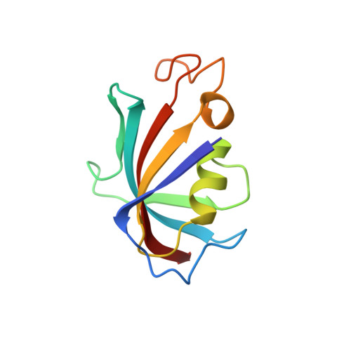
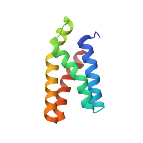
    2RSE

  • PubMed Abstract: 

    Pseudo contact shifts (PCSs) induced by paramagnetic lanthanide ions fixed in a protein frame provide long-range distance and angular information, and are valuable for the structure determination of protein-protein and protein-ligand complexes. We have been developing a lanthanide-binding peptide tag (hereafter LBT) anchored at two points via a peptide bond and a disulfide bond to the target proteins. However, the magnetic susceptibility tensor displays symmetry, which can cause multiple degenerated solutions in a structure calculation based solely on PCSs. Here we show a convenient method for resolving this degeneracy by changing the spacer length between the LBT and target protein. We applied this approach to PCS-based rigid body docking between the FKBP12-rapamycin complex and the mTOR FRB domain, and demonstrated that degeneracy could be resolved using the PCS restraints obtained from two-point anchored LBT with two different spacer lengths. The present strategy will markedly increase the usefulness of two-point anchored LBT for protein complex structure determination.


  • Organizational Affiliation
    • Department of Structural Biology, Faculty of Advanced Life Science, Hokkaido University, N-21, W-11, Sapporo 001-0021, Japan.

Macromolecule Content 

  • Total Structure Weight: 23.49 kDa 
  • Atom Count: 1,630 
  • Modeled Residue Count: 201 
  • Deposited Residue Count: 201 
  • Unique protein chains: 2

Macromolecules

Find similar proteins by:|  3D Structure
Entity ID: 1
MoleculeChains  Sequence LengthOrganismDetailsImage
Peptidyl-prolyl cis-trans isomerase FKBP1A107Homo sapiensMutation(s): 0 
Gene Names: FKBP1AFKBP1FKBP12
EC: 5.2.1.8
UniProt & NIH Common Fund Data Resources
Find proteins for P62942 (Homo sapiens)
Explore P62942 
Go to UniProtKB:  P62942
PHAROS:  P62942
GTEx:  ENSG00000088832 
Entity Groups
Sequence Clusters30% Identity50% Identity70% Identity90% Identity95% Identity100% Identity
UniProt GroupP62942
Sequence Annotations
Expand
Reference Sequence
Find similar proteins by:|  3D Structure
Entity ID: 2
MoleculeChains  Sequence LengthOrganismDetailsImage
Serine/threonine-protein kinase mTOR94Homo sapiensMutation(s): 0 
Gene Names: MTORFRAPFRAP1FRAP2RAFT1RAPT1
EC: 2.7.11.1 (PDB Primary Data), 2.7.10.2 (UniProt)
UniProt & NIH Common Fund Data Resources
Find proteins for P42345 (Homo sapiens)
Explore P42345 
Go to UniProtKB:  P42345
PHAROS:  P42345
GTEx:  ENSG00000198793 
Entity Groups
Sequence Clusters30% Identity50% Identity70% Identity90% Identity95% Identity100% Identity
UniProt GroupP42345
Sequence Annotations
Expand
Reference Sequence

Small Molecules

Experimental Data & Validation

Experimental Data

  • Method: SOLUTION NMR
  • Conformers Calculated: 100 
  • Conformers Submitted: 20 
  • Selection Criteria: structures with the lowest energy 

Structure Validation

View Full Validation Report


Entry History 

Deposition Data

Revision History  (Full details and data files)

  • Version 1.0: 2012-05-30
    Type: Initial release
  • Version 1.1: 2023-06-14
    Changes: Data collection, Database references, Other
  • Version 1.2: 2024-05-15
    Changes: Data collection, Database references