2RQ1 | pdb_00002rq1

Solution structure of the 4.1R FERM alpha lobe domain


Experimental Data Snapshot

  • Method: SOLUTION NMR
  • Conformers Calculated: 50 
  • Conformers Submitted: 20 
  • Selection Criteria: structures with the lowest energy 

wwPDB Validation 3D Report Full Report

Validation slider image for 2RQ1

This is version 1.3 of the entry. See complete history

Literature

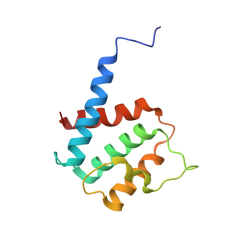
Solution structure and glycophorin C binding studies of the protein 4.1R FERM alpha-lobe domain

Kusunoki, H.Kohno, T.

(2009) Proteins 76: 255-260

Macromolecule Content 

  • Total Structure Weight: 12.42 kDa 
  • Atom Count: 870 
  • Modeled Residue Count: 109 
  • Deposited Residue Count: 109 
  • Unique protein chains: 1

Macromolecules

Find similar proteins by:|  3D Structure
Entity ID: 1
MoleculeChains  Sequence LengthOrganismDetailsImage
Protein 4.1109Homo sapiensMutation(s): 0 
Gene Names: EPB41E41P
UniProt & NIH Common Fund Data Resources
Find proteins for P11171 (Homo sapiens)
Explore P11171 
Go to UniProtKB:  P11171
PHAROS:  P11171
GTEx:  ENSG00000159023 
Entity Groups
Sequence Clusters30% Identity50% Identity70% Identity90% Identity95% Identity100% Identity
UniProt GroupP11171
Sequence Annotations
Expand
Reference Sequence

Experimental Data & Validation

Experimental Data

  • Method: SOLUTION NMR
  • Conformers Calculated: 50 
  • Conformers Submitted: 20 
  • Selection Criteria: structures with the lowest energy 

Structure Validation

View Full Validation Report


Entry History 

Deposition Data

Revision History  (Full details and data files)

  • Version 1.0: 2009-04-14
    Type: Initial release
  • Version 1.1: 2011-07-13
    Changes: Version format compliance
  • Version 1.2: 2022-03-16
    Changes: Data collection, Database references, Derived calculations
  • Version 1.3: 2024-05-29
    Changes: Data collection