2RNF

X-RAY CRYSTAL STRUCTURE OF HUMAN RIBONUCLEASE 4 IN COMPLEX WITH D(UP)


Experimental Data Snapshot

  • Method: X-RAY DIFFRACTION
  • Resolution: 2.40 Å
  • R-Value Free: 0.215 
  • R-Value Work: 0.160 
  • R-Value Observed: 0.160 

wwPDB Validation   3D Report Full Report


This is version 1.3 of the entry. See complete history


Literature

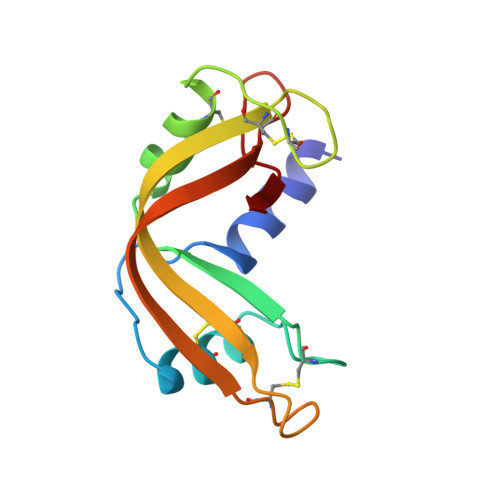
The three-dimensional structure of human RNase 4, unliganded and complexed with d(Up), reveals the basis for its uridine selectivity.

Terzyan, S.S.Peracaula, R.de Llorens, R.Tsushima, Y.Yamada, H.Seno, M.Gomis-Ruth, F.X.Coll, M.

(1999) J Mol Biol 285: 205-214

  • DOI: https://doi.org/10.1006/jmbi.1998.2288
  • Primary Citation of Related Structures:  
    1RNF, 2RNF

  • PubMed Abstract: 

    The RNase 4 family is unique among RNase enzymes, displaying the highest level of sequence similarity and encompassing the shortest polypeptide chain. It is the only one showing high specificity. The human representative is an intracellular and plasma enzyme, first isolated from colon adenocarcinoma cell line HT-29. The crystal structures of human recombinant RNase 4, unliganded and in complex with d(Up), have been determined, revealing in the unique active site an explanation for the uridine specificity. Arg101, at a position not involved in catalysis in the other RNase enzymes, penetrates the enzyme moiety shaping the recognition pocket, a flip that is mediated by the interaction with the (shorter chain) C-terminal carboxylate group, providing an anchoring point for the O4 atom of the substrate uridine. The bulky Phe42 side-chain forces Asp80 to be in the chi1=-72.49 degrees rotamer, accepting a hydrogen bond from Thr44, further converting the latter into a hydrogen bond acceptor. This favours an interaction with the -NH-donor group of uridine at position 3 over that with the =N-acceptor of cytidine. The two chemical groups that distinguish uracyl from cytosine are used by the enzyme to discriminate between these two bases.


  • Organizational Affiliation

    Centre d'Investigació i Desenvolupament, C.S.I.C., Jordi Girona, 18-26, Barcelona, 08034, Spain.


Macromolecules
Find similar proteins by:  (by identity cutoff)  |  3D Structure
Entity ID: 1
MoleculeChains Sequence LengthOrganismDetailsImage
RIBONUCLEASE 4
A, B
120Homo sapiensMutation(s): 0 
EC: 3.1.27
UniProt & NIH Common Fund Data Resources
Find proteins for P34096 (Homo sapiens)
Explore P34096 
Go to UniProtKB:  P34096
PHAROS:  P34096
GTEx:  ENSG00000258818 
Entity Groups  
Sequence Clusters30% Identity50% Identity70% Identity90% Identity95% Identity100% Identity
UniProt GroupP34096
Sequence Annotations
Expand
  • Reference Sequence
Small Molecules
Ligands 1 Unique
IDChains Name / Formula / InChI Key2D Diagram3D Interactions
UM3
Query on UM3

Download Ideal Coordinates CCD File 
C [auth A],
D [auth B]
2'-DEOXYURIDINE 3'-MONOPHOSPHATE
C9 H13 N2 O8 P
LXKGKXYIAAKOCT-SHYZEUOFSA-N
Experimental Data & Validation

Experimental Data

  • Method: X-RAY DIFFRACTION
  • Resolution: 2.40 Å
  • R-Value Free: 0.215 
  • R-Value Work: 0.160 
  • R-Value Observed: 0.160 
  • Space Group: P 1 21 1
Unit Cell:
Length ( Å )Angle ( ˚ )
a = 32.83α = 90
b = 94.33β = 107.2
c = 40.66γ = 90
Software Package:
Software NamePurpose
DENZOdata reduction
SCALEPACKdata scaling
AMoREphasing
X-PLORrefinement

Structure Validation

View Full Validation Report


Entry History 

Deposition Data

Revision History  (Full details and data files)

  • Version 1.0: 1999-11-10
    Type: Initial release
  • Version 1.1: 2008-04-26
    Changes: Version format compliance
  • Version 1.2: 2011-07-13
    Changes: Version format compliance
  • Version 1.3: 2023-08-30
    Changes: Data collection, Database references, Derived calculations, Refinement description