Crystal structure of a pH-stabilized mutant of villin headpiece.
Meng, J., McKnight, C.J.(2008) Biochemistry 47: 4644-4650
- PubMed: 18370407 Search on PubMed
- DOI: https://doi.org/10.1021/bi7022738
- Primary Citation Related Structures:
2RJV, 2RJY - PubMed Abstract:
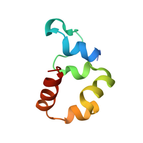
Villin-type headpiece domains are compact F-actin-binding motifs that have been used extensively as a model system to investigate protein folding by both experimental and computational methods. Villin headpiece (HP67) harbors a highly helical, thermostable, and autonomously folding subdomain in the C terminus (HP35), and because of this feature, HP67 is usually considered to be composed of a N- and C-terminal subdomain. Unlike the C-terminal subdomain, the N-terminal subdomain consists mainly of loops and turns, and the folding is dependent upon the presence of the C-terminal subdomain. The pH sensitivity of this subdomain is thought to arise from, at least partially, protonation of H41 buried in the hydrophobic core. Substitution of this histidine with tyrosine, another permissive residue at this position for naturally occurring sequences, increases not only the pH stability of HP67 but also the thermal stability and the cooperativity of thermal unfolding over a wide pH range (0.9-7.5). The crystal structures of wild-type HP67 and the H41Y mutant, determined under the same conditions, indicate that the H41Y substitution causes only localized rearrangement around the mutated residue. The F-actin-binding motif remains essentially the same after the mutation, accounting for the negligible effect of the mutation on F-actin affinity. The hydrogen bond formed between the imidazole ring of H41 and the backbone carbonyl of E14 of HP67 is eliminated by the H41Y mutation, which renders the extreme N terminus of H41Y more mobile; the hydrogen bond formed between the imidazole ring of H41 and the backbone nitrogen of D34 is replaced with that between the hydroxyl group of Y41 and the backbone nitrogen of D34 after the H41Y substitution. The increased hydrophobicity of tyrosine compensates for the loss of hydrogen bonds in the extreme N terminus and accounts for the increased stability and cooperativity of the H41Y mutant.
- Department of Physiology and Biophysics, Boston University School of Medicine, Boston, MA 02118, USA.
Organizational Affiliation: