2RJR

Substrate mimic bound to SgTAM


Experimental Data Snapshot

  • Method: X-RAY DIFFRACTION
  • Resolution: 2.10 Å
  • R-Value Free: 0.212 
  • R-Value Work: 0.173 
  • R-Value Observed: 0.176 

wwPDB Validation   3D Report Full Report


Ligand Structure Quality Assessment 


This is version 2.0 of the entry. See complete history


Literature

Design and characterization of mechanism-based inhibitors for the tyrosine aminomutase SgTAM.

Montavon, T.J.Christianson, C.V.Festin, G.M.Shen, B.Bruner, S.D.

(2008) Bioorg Med Chem Lett 18: 3099-3102

  • DOI: https://doi.org/10.1016/j.bmcl.2007.11.046
  • Primary Citation of Related Structures:  
    2QVE, 2RJR, 2RJS

  • PubMed Abstract: 

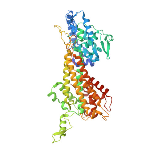
    The synthesis and evaluation of two classes of inhibitors for SgTAM, a 4-methylideneimidazole-5-one (MIO) containing tyrosine aminomutase, are described. A mechanism-based strategy was used to design analogs that mimic the substrate or product of the reaction and form covalent interactions with the enzyme through the MIO prosthetic group. The analogs were characterized by measuring inhibition constants and X-ray crystallographic structural analysis of the co-complexes bound to the aminomutase, SgTAM.


  • Organizational Affiliation

    Department of Chemistry, Merkert Chemistry Center, Boston College, Chestnut Hill, MA 02467, USA.


Macromolecules
Find similar proteins by:  (by identity cutoff)  |  3D Structure
Entity ID: 1
MoleculeChains Sequence LengthOrganismDetailsImage
Tyrosine aminomutase
A, B
537Streptomyces globisporusMutation(s): 0 
Gene Names: SgcC4
EC: 5.4.3.6
UniProt
Find proteins for Q8GMG0 (Streptomyces globisporus)
Explore Q8GMG0 
Go to UniProtKB:  Q8GMG0
Entity Groups  
Sequence Clusters30% Identity50% Identity70% Identity90% Identity95% Identity100% Identity
UniProt GroupQ8GMG0
Sequence Annotations
Expand
  • Reference Sequence
Small Molecules
Ligands 1 Unique
IDChains Name / Formula / InChI Key2D Diagram3D Interactions
295
Query on 295

Download Ideal Coordinates CCD File 
C [auth A](2S,3S)-3-(4-fluorophenyl)-2,3-dihydroxypropanoic acid
C9 H9 F O4
DWYLYIVEFVSGCP-YUMQZZPRSA-N
Modified Residues  1 Unique
IDChains TypeFormula2D DiagramParent
MDO
Query on MDO
A, B
L-PEPTIDE LINKINGC8 H11 N3 O3ALA, SER, GLY
Binding Affinity Annotations 
IDSourceBinding Affinity
295 PDBBind:  2RJR IC50: 1.16e+7 (nM) from 1 assay(s)
Experimental Data & Validation

Experimental Data

  • Method: X-RAY DIFFRACTION
  • Resolution: 2.10 Å
  • R-Value Free: 0.212 
  • R-Value Work: 0.173 
  • R-Value Observed: 0.176 
  • Space Group: P 21 21 2
Unit Cell:
Length ( Å )Angle ( ˚ )
a = 92.245α = 90
b = 145.648β = 90
c = 75.114γ = 90
Software Package:
Software NamePurpose
DENZOdata reduction
SCALEPACKdata scaling
CNSrefinement
PDB_EXTRACTdata extraction
HKL-2000data collection
CNSphasing

Structure Validation

View Full Validation Report


Ligand Structure Quality Assessment 


Entry History 

Deposition Data

Revision History  (Full details and data files)

  • Version 1.0: 2008-01-15
    Type: Initial release
  • Version 1.1: 2011-07-13
    Changes: Version format compliance
  • Version 1.2: 2011-11-16
    Changes: Atomic model
  • Version 1.3: 2017-10-25
    Changes: Refinement description
  • Version 1.4: 2023-08-30
    Changes: Data collection, Database references, Derived calculations, Refinement description
  • Version 2.0: 2023-11-15
    Changes: Atomic model, Data collection, Derived calculations