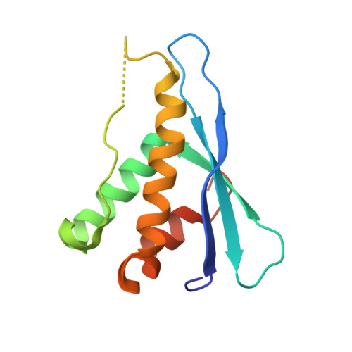
Crystal structures of PI3K-C2alpha PX domain indicate conformational change associated with ligand binding
Parkinson, G.N., Vines, D., Driscoll, P.C., Djordjevic, S.(2008) BMC Struct Biol 8: 13-13
- PubMed: 18312637 Search on PubMedSearch on PubMed Central
- DOI: https://doi.org/10.1186/1472-6807-8-13
- Primary Citation Related Structures:
2REA, 2RED - PubMed Abstract:
PX domains have specialized protein structures involved in binding of phosphoinositides (PIs). Through binding to the various PIs PX domains provide site-specific membrane signals to modulate the intracellular localisation and biological activity of effector proteins. Several crystal structures of these domains are now available from a variety of proteins. All PX domains contain a canonical core structure with main differences exhibited within the loop regions forming the phosphoinositide binding pockets. It is within these areas that the molecular basis for ligand specificity originates. We now report two new structures of PI3K-C2alpha PX domain that crystallised in a P3121 space group. The two structures, refined to 2.1 A and 2.5 A, exhibit significantly different conformations of the phosphoinositide-binding loops. Unexpectedly, in one of the structures, we have detected a putative-ligand trapped in the binding site during the process of protein purification and crystallisation. The two structures reported here provide a more complete description of the phosphoinositide binding region compared to the previously reported 2.6 A crystal structure of human PI3K-C2alpha PX where this region was highly disordered. The structures enabled us to further analyse PI specificity and to postulate that the observed conformational change could be related to ligand-binding.
- Cancer Research UK Biomolecular Structure Group, The School of Pharmacy, University of London, 29-39 Brunswick Square, London WC1N 1AX, UK. gary.parkinson@pharmacy.ac.uk
Organizational Affiliation: