Strain and near attack conformers in enzymic thiamin catalysis: X-ray crystallographic snapshots of bacterial transketolase in covalent complex with donor ketoses xylulose 5-phosphate and fructose 6-phosphate, and in noncovalent complex with acceptor aldose ribose 5-phosphate.
Asztalos, P., Parthier, C., Golbik, R., Kleinschmidt, M., Hubner, G., Weiss, M.S., Friedemann, R., Wille, G., Tittmann, K.(2007) Biochemistry 46: 12037-12052
- PubMed: 17914867 Search on PubMed
- DOI: https://doi.org/10.1021/bi700844m
- Primary Citation Related Structures:
2R5N, 2R8O, 2R8P - PubMed Abstract:
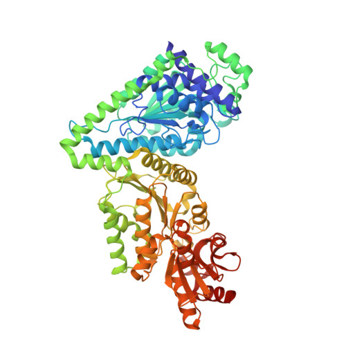
Transketolase is a prominent thiamin diphosphate-dependent enzyme in sugar metabolism that catalyzes the reversible transfer of a 2-carbon dihydroxyethyl fragment between a donor ketose and an acceptor aldose. The X-ray structures of transketolase from E. coli in a covalent complex with donor ketoses d-xylulose 5-phosphate (X5P) and d-fructose 6-phosphate (F6P) at 1.47 A and 1.65 A resolution reveal significant strain in the tetrahedral cofactor-sugar adducts with a 25-30 degrees out-of-plane distortion of the C2-Calpha bond connecting the substrates' carbonyl with the C2 of the cofactor's thiazolium part. Both intermediates adopt very similar extended conformations in the active site with a perpendicular orientation of the scissile C2-C3 sugar bond relative to the thiazolium ring. The sugar-derived hydroxyl groups of the intermediates form conserved hydrogen bonds with one Asp side chain, with a cluster of His residues and with the N4' of the aminopyrimidine ring of the cofactor. The phosphate moiety is held in place by electrostatic and hydrogen-bonding interactions with Arg, His, and Ser side chains. With the exception of the thiazolium part of the cofactor, no structural changes are observable during intermediate formation indicating that the active site is poised for catalysis. DFT calculations on both X5P-thiamin and X5P-thiazolium models demonstrate that an out-of-plane distortion of the C2-Calpha bond is energetically more favorable than a coplanar bond. The X-ray structure with the acceptor aldose d-ribose 5-phosphate (R5P) noncovalently bound in the active site suggests that the sugar is present in multiple forms: in a strained ring-closed beta-d-furanose form in C2-exo conformation as well as in an extended acyclic aldehyde form, with the reactive C1 aldo function held close to Calpha of the presumably planar carbanion/enamine intermediate. The latter form of R5P may be viewed as a near attack conformation. The R5P binding site overlaps with those of the leaving group moieties of the covalent donor-cofactor adducts, demonstrating that R5P directly competes with the donor-derived products glyceraldehyde 3-phosphate and erythrose 4-phosphate, which are substrates of the reverse reaction, for the same docking site at the active site and reaction with the DHEThDP enamine.
- Institut für Biochemie/Biotechnologie, Martin-Luther-University Halle-Wittenberg, Kurt-Mothes-Strasse 3, 06120 Halle/Saale, Germany.
Organizational Affiliation: