2R5L

Crystal structure of lactoperoxidase at 2.4A resolution


Experimental Data Snapshot

  • Method: X-RAY DIFFRACTION
  • Resolution: 2.40 Å
  • R-Value Free: 0.203 
  • R-Value Work: 0.196 
  • R-Value Observed: 0.196 

wwPDB Validation   3D Report Full Report


Ligand Structure Quality Assessment 


This is version 2.1 of the entry. See complete history


Literature

Crystal Structure of Lactoperoxidase at 2.4 A Resolution.

Singh, A.K.Singh, N.Sharma, S.Singh, S.B.Kaur, P.Bhushan, A.Srinivasan, A.Singh, T.P.

(2007) J Mol Biol 376: 1060-1075

  • DOI: https://doi.org/10.1016/j.jmb.2007.12.012
  • Primary Citation of Related Structures:  
    2R5L

  • PubMed Abstract: 

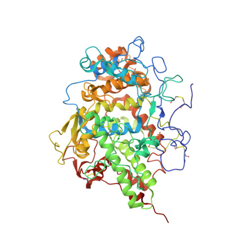
    Lactoperoxidase (LPO) is a member of the mammalian peroxidase superfamily. It catalyzes the oxidation of thiocyanate and halides. Freshly isolated and purified samples of caprine LPO were saturated with ammonium iodide and crystallized using 20% polyethylene glycol 3350 in a hanging drop vapor diffusion setup. The structure has been determined using X-ray crystallographic method and refined to R(cryst) and R(free) factors of 0.196 and 0.203, respectively. The structure determination revealed an unexpected phosphorylation of Ser198 in LPO, which is also confirmed by anti-phosphoserine antibody binding studies. The structure is also notable for observing densities for glycan chains at all the four potential glycosylation sites. Caprine LPO consists of a single polypeptide chain of 595 amino acid residues and folds into an oval-shaped structure. The structure contains 20 well-defined alpha-helices of varying lengths including a helix, H(2a), unique to LPO, and two short antiparallel beta-strands. The structure confirms that the heme group is covalently linked to the protein through two ester linkages involving carboxylic groups of Glu258 and Asp108 and modified methyl groups of pyrrole rings A and C, respectively. The heme moiety is slightly distorted from planarity, but pyrrole ring B is distorted considerably. However, an iron atom is displaced only by 0.1 A from the plane of the heme group toward the proximal site. The substrate diffusing channel in LPO is cylindrical in shape with a diameter of approximately 6 A. Two histidine residues and six buried water molecules are connected through a hydrogen-bonded chain from the distal heme cavity to the surface of protein molecule and seemingly form the basis of proton relay for catalytic action. Ten iodide ions have been observed in the structure. Out of these, only one iodide ion is located in the distal heme cavity and is hydrogen bonded to the water molecule W1. W1 is also hydrogen bonded to the heme iron as well as to distal His109. The structure contains a calcium ion that is coordinated to seven oxygen atoms and forms a typical pentagonal bipyramidal coordination geometry.


  • Organizational Affiliation

    Department of Biophysics, All India Institute of Medical Sciences, Ansari Nagar, New Delhi - 110 029, India.


Macromolecules
Find similar proteins by:  (by identity cutoff)  |  3D Structure
Entity ID: 1
MoleculeChains Sequence LengthOrganismDetailsImage
Lactoperoxidase595Capra hircusMutation(s): 0 
EC: 1.11.1.7
UniProt
Find proteins for A0A452E9Y6 (Capra hircus)
Explore A0A452E9Y6 
Go to UniProtKB:  A0A452E9Y6
Entity Groups  
Sequence Clusters30% Identity50% Identity70% Identity90% Identity95% Identity100% Identity
UniProt GroupA0A452E9Y6
Sequence Annotations
Expand
  • Reference Sequence
Oligosaccharides

Help

Entity ID: 2
MoleculeChains Length2D Diagram Glycosylation3D Interactions
alpha-D-mannopyranose-(1-4)-2-acetamido-2-deoxy-beta-D-glucopyranose-(1-4)-2-acetamido-2-deoxy-beta-D-glucopyranose
B, C, D
3N-Glycosylation
Glycosylation Resources
GlyTouCan:  G62182OO
GlyCosmos:  G62182OO
GlyGen:  G62182OO
Entity ID: 3
MoleculeChains Length2D Diagram Glycosylation3D Interactions
2-acetamido-2-deoxy-beta-D-glucopyranose-(1-4)-2-acetamido-2-deoxy-beta-D-glucopyranose
E
2N-Glycosylation
Glycosylation Resources
GlyTouCan:  G42666HT
GlyCosmos:  G42666HT
GlyGen:  G42666HT
Small Molecules
Ligands 3 Unique
IDChains Name / Formula / InChI Key2D Diagram3D Interactions
HEM
Query on HEM

Download Ideal Coordinates CCD File 
Q [auth A]PROTOPORPHYRIN IX CONTAINING FE
C34 H32 Fe N4 O4
KABFMIBPWCXCRK-RGGAHWMASA-L
IOD
Query on IOD

Download Ideal Coordinates CCD File 
G [auth A]
H [auth A]
I [auth A]
J [auth A]
K [auth A]
G [auth A],
H [auth A],
I [auth A],
J [auth A],
K [auth A],
L [auth A],
M [auth A],
N [auth A],
O [auth A],
P [auth A]
IODIDE ION
I
XMBWDFGMSWQBCA-UHFFFAOYSA-M
CA
Query on CA

Download Ideal Coordinates CCD File 
F [auth A]CALCIUM ION
Ca
BHPQYMZQTOCNFJ-UHFFFAOYSA-N
Modified Residues  1 Unique
IDChains TypeFormula2D DiagramParent
SEP
Query on SEP
A
L-PEPTIDE LINKINGC3 H8 N O6 PSER
Experimental Data & Validation

Experimental Data

  • Method: X-RAY DIFFRACTION
  • Resolution: 2.40 Å
  • R-Value Free: 0.203 
  • R-Value Work: 0.196 
  • R-Value Observed: 0.196 
  • Space Group: P 1 21 1
Unit Cell:
Length ( Å )Angle ( ˚ )
a = 54.19α = 90
b = 80.81β = 102.95
c = 77.04γ = 90
Software Package:
Software NamePurpose
CNSrefinement
MAR345dtbdata collection
DENZOdata reduction
SCALEPACKdata scaling
AMoREphasing

Structure Validation

View Full Validation Report


Ligand Structure Quality Assessment 


Entry History 

Deposition Data

Revision History  (Full details and data files)

  • Version 1.0: 2007-09-18
    Type: Initial release
  • Version 1.1: 2011-07-13
    Changes: Non-polymer description, Version format compliance
  • Version 2.0: 2020-07-29
    Type: Remediation
    Reason: Carbohydrate remediation
    Changes: Atomic model, Data collection, Derived calculations, Structure summary
  • Version 2.1: 2023-10-25
    Changes: Data collection, Database references, Refinement description, Structure summary