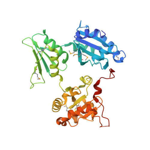
2R2J

crystal structure of human ERp44


Experimental Data Snapshot

  • Method: X-RAY DIFFRACTION
  • Resolution: 2.60 Å
  • R-Value Free: 0.263 
  • R-Value Work: 0.217 
  • R-Value Observed: 0.219 

wwPDB Validation   3D Report Full Report


This is version 1.1 of the entry. See complete history


Literature

Crystal structure of human ERp44 shows a dynamic functional modulation by its carboxy-terminal tail.

Wang, L.K.Wang, L.Vavassori, S.Li, S.J.Ke, H.Anelli, T.Degano, M.Ronzoni, R.Sitia, R.Sun, F.Wang, C.C.

(2008) EMBO Rep 

  • DOI: https://doi.org/10.1038/embor.2008.88
  • Primary Citation of Related Structures:  
    2R2J

  • PubMed Abstract: 

    ERp44 mediates thiol-dependent retention in the early secretory pathway, forming mixed disulphides with substrate proteins through its conserved CRFS motif. Here, we present its crystal structure at a resolution of 2.6 A. Three thioredoxin domains-a, b and b'-are arranged in a clover-like structure. A flexible carboxy-terminal tail turns back to the b' and a domains, shielding a hydrophobic pocket in domain b' and a hydrophobic patch around the CRFS motif in domain a. Mutational and functional studies indicate that the C-terminal tail gates the CRFS area and the adjacent hydrophobic pocket, dynamically regulating protein quality control.


  • Organizational Affiliation

    National Laboratory of Biomacromolecules, Institute of Biophysics, Chinese Academy of Sciences, Beijing 100101, China.


Macromolecules
Find similar proteins by:  (by identity cutoff)  |  3D Structure
Entity ID: 1
MoleculeChains Sequence LengthOrganismDetailsImage
Thioredoxin domain-containing protein 4382Homo sapiensMutation(s): 0 
Gene Names: TXNDC4ERP44KIAA0573
UniProt & NIH Common Fund Data Resources
Find proteins for Q9BS26 (Homo sapiens)
Explore Q9BS26 
Go to UniProtKB:  Q9BS26
PHAROS:  Q9BS26
GTEx:  ENSG00000023318 
Entity Groups  
Sequence Clusters30% Identity50% Identity70% Identity90% Identity95% Identity100% Identity
UniProt GroupQ9BS26
Sequence Annotations
Expand
  • Reference Sequence
Small Molecules
Modified Residues  1 Unique
IDChains TypeFormula2D DiagramParent
MSE
Query on MSE
A
L-PEPTIDE LINKINGC5 H11 N O2 SeMET
Experimental Data & Validation

Experimental Data

  • Method: X-RAY DIFFRACTION
  • Resolution: 2.60 Å
  • R-Value Free: 0.263 
  • R-Value Work: 0.217 
  • R-Value Observed: 0.219 
  • Space Group: P 31 2 1
Unit Cell:
Length ( Å )Angle ( ˚ )
a = 83.501α = 90
b = 83.501β = 90
c = 123.137γ = 120
Software Package:
Software NamePurpose
REFMACrefinement
HKL-2000data collection
HKL-2000data reduction
HKL-2000data scaling
SOLVEphasing

Structure Validation

View Full Validation Report


Entry History 

Deposition Data

Revision History  (Full details and data files)

  • Version 1.0: 2008-07-08
    Type: Initial release
  • Version 1.1: 2011-07-13
    Changes: Advisory, Version format compliance