


Crystal structure of a kinase MARK2/Par-1 mutant
Panneerselvam, S., Marx, A., Mandelkow, E.-M., Mandelkow, E.To be published.
Experimental Data Snapshot
Starting Model: experimental
View more details
wwPDB Validation 3D Report Full Report

Entity ID: 1 | |||||
|---|---|---|---|---|---|
| Molecule | Chains | Sequence Length | Organism | Details | Image |
| Serine/threonine-protein kinase MARK2 | 327 | Rattus norvegicus | Mutation(s): 1 Gene Names: MARK2 EC: 2.7.11.1 (PDB Primary Data), 2.7.11.26 (UniProt) | 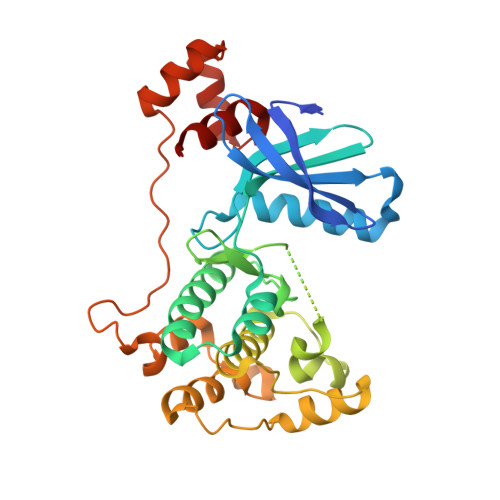 | |
UniProt | |||||
Entity Groups | |||||
| Sequence Clusters | 30% Identity50% Identity70% Identity90% Identity95% Identity100% Identity | ||||
| UniProt Group | O08679 | ||||
Sequence AnnotationsExpand | |||||
Reference Sequence | |||||
| Length ( Å ) | Angle ( ˚ ) |
|---|---|
| a = 120.405 | α = 90 |
| b = 120.405 | β = 90 |
| c = 99.919 | γ = 120 |
| Software Name | Purpose |
|---|---|
| DENZO | data reduction |
| SCALEPACK | data scaling |
| PHASER | phasing |
| REFMAC | refinement |
| PDB_EXTRACT | data extraction |


