2QVW | pdb_00002qvw

Structure of Giardia Dicer refined against twinned data


Experimental Data Snapshot

  • Method: X-RAY DIFFRACTION
  • Resolution: 3.00 Å
  • R-Value Free: 
    0.263 (Depositor), 0.246 (DCC) 
  • R-Value Work: 
    0.221 (Depositor), 0.216 (DCC) 

Starting Model: experimental
View more details

wwPDB Validation 3D Report Full Report

Validation slider image for 2QVW

This is version 1.3 of the entry. See complete history

Literature

An unusual case of pseudo-merohedral twinning in orthorhombic crystals of Dicer

Macrae, I.J.Doudna, J.A.

(2007) Acta Crystallogr D Biol Crystallogr 63: 993-999

  • DOI: https://doi.org/10.1107/S0907444907036128
  • Primary Citation Related Structures: 
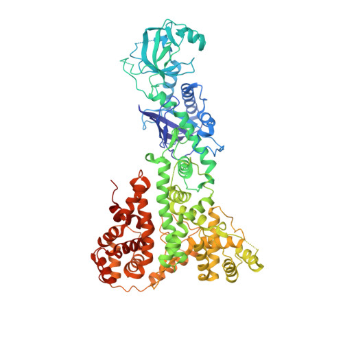
    2QVW

  • PubMed Abstract: 

    The crystal structure of the enzyme Dicer from Giardia intestinalis was solved to 3.3 A resolution by MAD using crystals belonging to space group P2(1)2(1)2 [Macrae et al. (2006), Science, 311, 195-198]. These crystals were derived from crystals that diffracted X-rays to 3.0 A resolution but were refractory to structure determination because they were twinned. It is shown here that the original Dicer crystals represent an unusual case of perfect pseudo-merohedral twinning of orthorhombic crystals. Before the twinning problem was overcome, it was possible to calculate a low-resolution electron-density map in space group P4(1) that was used to build a partial molecular model. Experimental phases were sufficient to identify heavy-atom sites that indicated space-group inconsistency, leading to identification of the true space group. This information guided the search for different crystallization conditions that yielded untwinned crystals and ultimately a fully interpretable electron-density map.


  • Organizational Affiliation
    • Howard Hughes Medical Institute, Department of Molecular and Cell Biology, University of California, Berkeley, CA 94720, USA.

Macromolecule Content 

  • Total Structure Weight: 332.9 kDa 
  • Atom Count: 22,372 
  • Modeled Residue Count: 2,896 
  • Deposited Residue Count: 3,024 
  • Unique protein chains: 1

Macromolecules

Find similar proteins by:|  3D Structure
Entity ID: 1
MoleculeChains  Sequence LengthOrganismDetailsImage
GLP_546_48378_50642
A, B, C, D
756Giardia duodenalisMutation(s): 0 
EC: 3.1.26.3 (PDB Primary Data), 3.1.26 (UniProt)
UniProt
Find proteins for A8BQJ3 (Giardia intestinalis (strain ATCC 50803 / WB clone C6))
Explore A8BQJ3 
Go to UniProtKB:  A8BQJ3
Entity Groups
Sequence Clusters30% Identity50% Identity70% Identity90% Identity95% Identity100% Identity
UniProt GroupA8BQJ3
Sequence Annotations
Expand
Reference Sequence

Small Molecules

Ligands 1 Unique
IDChains Name / Formula / InChI Key2D Diagram3D Interactions
MN

Query on MN


Download:Ideal Coordinates CCD File
AA [auth C]
BA [auth C]
CA [auth C]
DA [auth C]
E [auth A]
AA [auth C],
BA [auth C],
CA [auth C],
DA [auth C],
E [auth A],
EA [auth C],
F [auth A],
FA [auth C],
G [auth A],
GA [auth C],
H [auth A],
HA [auth C],
I [auth A],
IA [auth D],
J [auth A],
JA [auth D],
K [auth A],
KA [auth D],
L [auth A],
LA [auth D],
M [auth A],
MA [auth D],
N [auth A],
NA [auth D],
O [auth A],
OA [auth D],
P [auth A],
PA [auth D],
Q [auth B],
QA [auth D],
R [auth B],
S [auth B],
T [auth B],
U [auth B],
V [auth B],
W [auth B],
X [auth B],
Y [auth B],
Z [auth C]
MANGANESE (II) ION
Mn
WAEMQWOKJMHJLA-UHFFFAOYSA-N

Experimental Data & Validation

Experimental Data

  • Method: X-RAY DIFFRACTION
  • Resolution: 3.00 Å
  • R-Value Free:  0.263 (Depositor), 0.246 (DCC) 
  • R-Value Work:  0.221 (Depositor), 0.216 (DCC) 
Space Group: P 21 21 2
Unit Cell:
Length ( Å )Angle ( ˚ )
a = 155.409α = 90
b = 173.49β = 90
c = 155.453γ = 90
Software Package:
Software NamePurpose
CNSrefinement
PDB_EXTRACTdata extraction
HKL-2000data collection
HKL-2000data reduction
HKL-2000data scaling
CNSphasing

Structure Validation

View Full Validation Report


Entry History 

Deposition Data

Revision History  (Full details and data files)

  • Version 1.0: 2007-09-25
    Type: Initial release
  • Version 1.1: 2011-07-13
    Changes: Version format compliance
  • Version 1.2: 2018-01-24
    Changes: Structure summary
  • Version 1.3: 2023-08-30
    Changes: Data collection, Database references, Derived calculations, Refinement description