2QUD | pdb_00002qud

PP7 Coat Protein Dimer


Experimental Data Snapshot

  • Method: X-RAY DIFFRACTION
  • Resolution: 1.60 Å
  • R-Value Free: 
    0.197 (Depositor), 0.208 (DCC) 
  • R-Value Work: 
    0.157 (Depositor), 0.174 (DCC) 
  • R-Value Observed: 
    0.159 (Depositor) 

Starting Model: experimental
View more details

wwPDB Validation 3D Report Full Report

Validation slider image for 2QUD

This is version 1.3 of the entry. See complete history

Literature

Structural basis for the coevolution of a viral RNA-protein complex.

Chao, J.A.Patskovsky, Y.Almo, S.C.Singer, R.H.

(2008) Nat Struct Mol Biol 15: 103-105

  • DOI: https://doi.org/10.1038/nsmb1327
  • Primary Citation Related Structures: 
    2QUD, 2QUX

  • PubMed Abstract: 

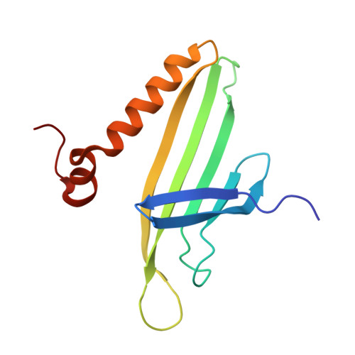
    The cocrystal structure of the PP7 bacteriophage coat protein in complex with its translational operator identifies a distinct mode of sequence-specific RNA recognition when compared to the well-characterized MS2 coat protein-RNA complex. The structure reveals the molecular basis of the PP7 coat protein's ability to selectively bind its cognate RNA, and it demonstrates that the conserved beta-sheet surface is a flexible architecture that can evolve to recognize diverse RNA hairpins.


  • Organizational Affiliation
    • Department of Anatomy, Albert Einstein College of Medicine, 1300 Morris Park Ave, Bronx, New York 10461, USA.

Macromolecule Content 

  • Total Structure Weight: 27.33 kDa 
  • Atom Count: 2,363 
  • Modeled Residue Count: 242 
  • Deposited Residue Count: 250 
  • Unique protein chains: 1

Macromolecules

Find similar proteins by:|  3D Structure
Entity ID: 1
MoleculeChains  Sequence LengthOrganismDetailsImage
Coat protein
A, B
125Pseudomonas phage PP7Mutation(s): 0 
Gene Names: PP7 Coat Protein
UniProt
Find proteins for P03630 (Pseudomonas phage PP7)
Explore P03630 
Go to UniProtKB:  P03630
Entity Groups
Sequence Clusters30% Identity50% Identity70% Identity90% Identity95% Identity100% Identity
UniProt GroupP03630
Sequence Annotations
Expand
Reference Sequence

Small Molecules

Ligands 1 Unique
IDChains Name / Formula / InChI Key2D Diagram3D Interactions
GOL

Query on GOL


Download:Ideal Coordinates CCD File
C [auth A]GLYCEROL
C3 H8 O3
PEDCQBHIVMGVHV-UHFFFAOYSA-N

Experimental Data & Validation

Experimental Data

  • Method: X-RAY DIFFRACTION
  • Resolution: 1.60 Å
  • R-Value Free:  0.197 (Depositor), 0.208 (DCC) 
  • R-Value Work:  0.157 (Depositor), 0.174 (DCC) 
  • R-Value Observed: 0.159 (Depositor) 
Space Group: H 3
Unit Cell:
Length ( Å )Angle ( ˚ )
a = 154.985α = 90
b = 154.985β = 90
c = 31.631γ = 120
Software Package:
Software NamePurpose
PHASERphasing
REFMACrefinement
HKL-2000data reduction
HKL-2000data scaling

Structure Validation

View Full Validation Report


Entry History 

Deposition Data

  • Released Date: 2007-12-18 
  • Deposition Author(s): Chao, J.A.

Revision History  (Full details and data files)

  • Version 1.0: 2007-12-18
    Type: Initial release
  • Version 1.1: 2011-07-13
    Changes: Non-polymer description, Version format compliance
  • Version 1.2: 2017-08-02
    Changes: Refinement description, Source and taxonomy
  • Version 1.3: 2023-08-30
    Changes: Data collection, Database references, Derived calculations, Refinement description