2QTW | pdb_00002qtw

The Crystal Structure of PCSK9 at 1.9 Angstroms Resolution Reveals structural homology to Resistin within the C-terminal domain


Experimental Data Snapshot

  • Method: X-RAY DIFFRACTION
  • Resolution: 1.90 Å
  • R-Value Free: 
    0.239 (Depositor), 0.244 (DCC) 
  • R-Value Work: 
    0.192 (Depositor), 0.197 (DCC) 
  • R-Value Observed: 
    0.194 (Depositor) 

wwPDB Validation 3D Report Full Report

Validation slider image for 2QTW

Ligand Structure Quality Assessment 


This is version 1.5 of the entry. See complete history

Literature

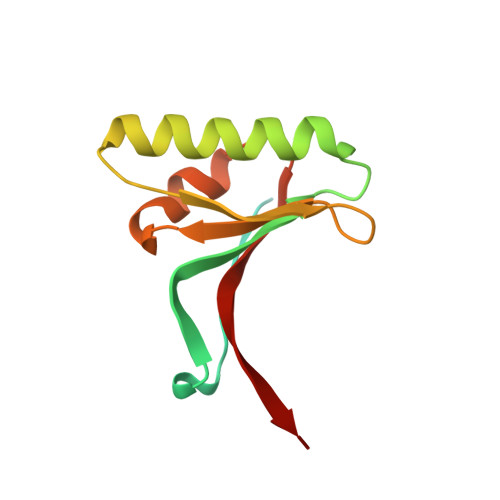
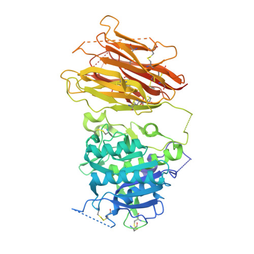
The self-inhibited structure of full-length PCSK9 at 1.9 A reveals structural homology with resistin within the C-terminal domain.

Hampton, E.N.Knuth, M.W.Li, J.Harris, J.L.Lesley, S.A.Spraggon, G.

(2007) Proc Natl Acad Sci U S A 104: 14604-14609

  • DOI: https://doi.org/10.1073/pnas.0703402104
  • Primary Citation Related Structures: 
    2QTW

  • PubMed Abstract: 

    Mutations in proprotein convertase subtilisin/kexin type 9 (PCSK9) are strongly associated with levels of low-density lipoprotein cholesterol in the blood plasma and, thereby, occurrence or resistance to atherosclerosis and coronary heart disease. Despite this importance, relatively little is known about the biology of PCSK9. Here, the crystal structure of a full-length construct of PCSK9 solved to 1.9-A resolution is presented. The structure contains a fully folded C-terminal cysteine-rich domain (CRD), showing a distinct structural similarity to the resistin homotrimer, a small cytokine associated with obesity and diabetes. This structural relationship between the CRD of PCSK9 and the resistin family is not observed in primary sequence comparisons and strongly suggests a distant evolutionary link between the two molecules. This three-dimensional homology provides insight into the function of PCSK9 at the molecular level and will help to dissect the link between PCSK9 and CHD.


  • Organizational Affiliation
    • Genomics Institute of the Novartis Research Foundation, 10675 John Jay Hopkins Drive, San Diego, CA 92121, USA.

Macromolecule Content 

  • Total Structure Weight: 72.55 kDa 
  • Atom Count: 4,697 
  • Modeled Residue Count: 578 
  • Deposited Residue Count: 670 
  • Unique protein chains: 2

Macromolecules

Find similar proteins by:|  3D Structure
Entity ID: 1
MoleculeChains  Sequence LengthOrganismDetailsImage
Proprotein convertase subtilisin/kexin type 9 Propeptide124Homo sapiensMutation(s): 0 
Gene Names: PCSK9NARC1
EC: 3.4.21
UniProt & NIH Common Fund Data Resources
Find proteins for Q8NBP7 (Homo sapiens)
Explore Q8NBP7 
Go to UniProtKB:  Q8NBP7
PHAROS:  Q8NBP7
GTEx:  ENSG00000169174 
Entity Groups
Sequence Clusters30% Identity50% Identity70% Identity90% Identity95% Identity100% Identity
UniProt GroupQ8NBP7
Sequence Annotations
Expand
Reference Sequence
Find similar proteins by:|  3D Structure
Entity ID: 2
MoleculeChains  Sequence LengthOrganismDetailsImage
Proprotein convertase subtilisin/kexin type 9546Homo sapiensMutation(s): 0 
Gene Names: PCSK9NARC1
EC: 3.4.21
UniProt & NIH Common Fund Data Resources
Find proteins for Q8NBP7 (Homo sapiens)
Explore Q8NBP7 
Go to UniProtKB:  Q8NBP7
PHAROS:  Q8NBP7
GTEx:  ENSG00000169174 
Entity Groups
Sequence Clusters30% Identity50% Identity70% Identity90% Identity95% Identity100% Identity
UniProt GroupQ8NBP7
Glycosylation
Glycosylation Sites: 1Go to GlyGen: Q8NBP7-1
Sequence Annotations
Expand
Reference Sequence

Small Molecules

Experimental Data & Validation

Experimental Data

  • Method: X-RAY DIFFRACTION
  • Resolution: 1.90 Å
  • R-Value Free:  0.239 (Depositor), 0.244 (DCC) 
  • R-Value Work:  0.192 (Depositor), 0.197 (DCC) 
  • R-Value Observed: 0.194 (Depositor) 
Space Group: P 21 21 21
Unit Cell:
Length ( Å )Angle ( ˚ )
a = 62.427α = 90
b = 70.149β = 90
c = 148.59γ = 90
Software Package:
Software NamePurpose
REFMACrefinement
HKL-2000data reduction
HKL-2000data scaling
PHASERphasing

Structure Validation

View Full Validation Report


Ligand Structure Quality Assessment 


Entry History 

Deposition Data

Revision History  (Full details and data files)

  • Version 1.0: 2007-09-18
    Type: Initial release
  • Version 1.1: 2011-07-13
    Changes: Non-polymer description, Version format compliance
  • Version 1.2: 2013-09-18
    Changes: Derived calculations
  • Version 1.3: 2020-02-05
    Changes: Data collection, Database references, Derived calculations, Source and taxonomy
  • Version 1.4: 2020-07-29
    Type: Remediation
    Reason: Carbohydrate remediation
    Changes: Data collection, Derived calculations, Structure summary
  • Version 1.5: 2024-10-16
    Changes: Data collection, Database references, Structure summary