


Crystal structure of a GTP-binding protein from the hyperthermophilic archaeon Sulfolobus solfataricus.
Wu, H., Sun, L., Brouns, S.J., Fu, S., Akerboom, J., Li, X., Zhang, C., Rao, Z., Van der Oost, J.To be published.
Experimental Data Snapshot
Entity ID: 1 | |||||
|---|---|---|---|---|---|
| Molecule | Chains | Sequence Length | Organism | Details | Image |
| GTP-binding protein | 364 | N/A | Mutation(s): 0 Gene Names: hflX, SSO0269 | 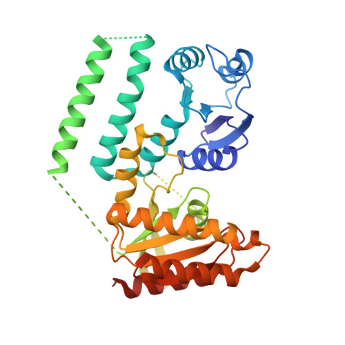 | |
UniProt | |||||
Entity Groups | |||||
| Sequence Clusters | 30% Identity50% Identity70% Identity90% Identity95% Identity100% Identity | ||||
| UniProt Group | Q980M3 | ||||
Sequence AnnotationsExpand | |||||
Reference Sequence | |||||
| Ligands 4 Unique | |||||
|---|---|---|---|---|---|
| ID | Chains | Name / Formula / InChI Key | 2D Diagram | 3D Interactions | |
| GDP Download:Ideal Coordinates CCD File | H [auth A] | GUANOSINE-5'-DIPHOSPHATE C10 H15 N5 O11 P2 QGWNDRXFNXRZMB-UUOKFMHZSA-N |  | ||
| CD Download:Ideal Coordinates CCD File | B [auth A], C [auth A], D [auth A], E [auth A] | CADMIUM ION Cd WLZRMCYVCSSEQC-UHFFFAOYSA-N |  | ||
| ACT Download:Ideal Coordinates CCD File | G [auth A], I [auth A], J [auth A] | ACETATE ION C2 H3 O2 QTBSBXVTEAMEQO-UHFFFAOYSA-M |  | ||
| MG Download:Ideal Coordinates CCD File | F [auth A] | MAGNESIUM ION Mg JLVVSXFLKOJNIY-UHFFFAOYSA-N |  | ||
| Length ( Å ) | Angle ( ˚ ) |
|---|---|
| a = 64.991 | α = 90 |
| b = 72.403 | β = 90 |
| c = 96.016 | γ = 90 |
| Software Name | Purpose |
|---|---|
| DENZO | data reduction |
| SCALEPACK | data scaling |
| REFMAC | refinement |
| PDB_EXTRACT | data extraction |
| CrystalClear | data collection |
| SHARP | phasing |
| DM | phasing |


