2QRY | pdb_00002qry

Periplasmic thiamin binding protein


Experimental Data Snapshot

  • Method: X-RAY DIFFRACTION
  • Resolution: 2.25 Å
  • R-Value Free: 
    0.241 (Depositor), 0.241 (DCC) 
  • R-Value Work: 
    0.198 (Depositor), 0.198 (DCC) 
  • R-Value Observed: 
    0.200 (Depositor) 

wwPDB Validation 3D Report Full Report

Validation slider image for 2QRY

Ligand Structure Quality Assessment 


This is version 1.2 of the entry. See complete history

Literature

Structural Similarities between Thiamin-Binding Protein and Thiaminase-I Suggest a Common Ancestor

Soriano, E.V.Rajashankar, K.R.Hanes, J.W.Bale, S.Begley, T.P.Ealick, S.E.

(2008) Biochemistry 47: 1346-1357

  • DOI: https://doi.org/10.1021/bi7018282
  • Primary Citation Related Structures: 
    2QRY

  • PubMed Abstract: 

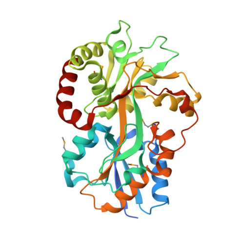
    ATP-binding cassette (ABC) transporters are responsible for the transport of a wide variety of water-soluble molecules and ions into prokaryotic cells. In Gram-negative bacteria, periplasmic-binding proteins deliver ions or molecules such as thiamin to the membrane-bound ABC transporter. The gene for the thiamin-binding protein tbpA has been identified in both Escherichia coli and Salmonella typhimurium. Here we report the crystal structure of TbpA from E. coli with bound thiamin monophosphate. The structure was determined at 2.25 A resolution using single-wavelength anomalous diffraction experiments, despite the presence of nonmerohedral twinning. The crystal structure shows that TbpA belongs to the group II periplasmic-binding protein family. Equilibrium binding measurements showed similar dissociation constants for thiamin, thiamin monophosphate, and thiamin pyrophosphate. Analysis of the binding site by molecular modeling demonstrated how TbpA binds all three forms of thiamin. A comparison of TbpA and thiaminase-I, a thiamin-degrading enzyme, revealed structural similarity between the two proteins, especially in domain 1, suggesting that the two proteins evolved from a common ancestor.


  • Organizational Affiliation
    • Department of Chemistry and Chemical Biology, Cornell University, Ithaca, New York 14853, USA.

Macromolecule Content 

  • Total Structure Weight: 148.68 kDa 
  • Atom Count: 10,279 
  • Modeled Residue Count: 1,245 
  • Deposited Residue Count: 1,320 
  • Unique protein chains: 1

Macromolecules

Find similar proteins by:|  3D Structure
Entity ID: 1
MoleculeChains  Sequence LengthOrganismDetailsImage
Thiamine-binding periplasmic protein
A, B, C, D
330Escherichia coliMutation(s): 0 
Gene Names: thiBtbpA
UniProt
Find proteins for P31550 (Escherichia coli (strain K12))
Explore P31550 
Go to UniProtKB:  P31550
Entity Groups
Sequence Clusters30% Identity50% Identity70% Identity90% Identity95% Identity100% Identity
UniProt GroupP31550
Sequence Annotations
Expand
Reference Sequence

Small Molecules

Experimental Data & Validation

Experimental Data

  • Method: X-RAY DIFFRACTION
  • Resolution: 2.25 Å
  • R-Value Free:  0.241 (Depositor), 0.241 (DCC) 
  • R-Value Work:  0.198 (Depositor), 0.198 (DCC) 
  • R-Value Observed: 0.200 (Depositor) 
Space Group: P 1 21 1
Unit Cell:
Length ( Å )Angle ( ˚ )
a = 69.04α = 90
b = 118.07β = 93.74
c = 90.33γ = 90
Software Package:
Software NamePurpose
CNSrefinement
HKL-2000data collection
HKL-2000data reduction
HKL-2000data scaling
MLPHAREphasing

Structure Validation

View Full Validation Report


Ligand Structure Quality Assessment 


Entry History 

Deposition Data

Revision History  (Full details and data files)

  • Version 1.0: 2008-02-05
    Type: Initial release
  • Version 1.1: 2011-07-13
    Changes: Version format compliance
  • Version 1.2: 2024-11-13
    Changes: Data collection, Database references, Derived calculations, Structure summary