2QRT | pdb_00002qrt

Crystal Structure of a disulfide trapped single chain trimer composed of the MHC I heavy chain H-2Kb Y84C, beta-2microglobulin, and ovalbumin-derived peptide.


Experimental Data Snapshot

  • Method: X-RAY DIFFRACTION
  • Resolution: 1.80 Å
  • R-Value Free: 
    0.239 (Depositor), 0.238 (DCC) 
  • R-Value Work: 
    0.208 (Depositor), 0.207 (DCC) 

Starting Model: experimental
View more details

wwPDB Validation 3D Report Full Report

Validation slider image for 2QRT

This is version 1.5 of the entry. See complete history

Literature

Structural engineering of pMHC reagents for T cell vaccines and diagnostics.

Mitaksov, V.Truscott, S.M.Lybarger, L.Connolly, J.M.Hansen, T.H.Fremont, D.H.

(2007) Chem Biol 14: 909-922

  • DOI: https://doi.org/10.1016/j.chembiol.2007.07.010
  • Primary Citation Related Structures: 
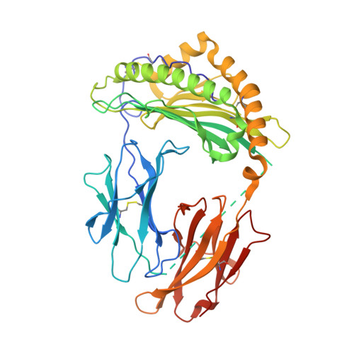
    2QRI, 2QRS, 2QRT

  • PubMed Abstract: 

    MHC class I peptide complexes (pMHC) are routinely used to enumerate T cell populations and are currently being evaluated as vaccines to tumors and specific pathogens. Herein, we describe the structures of three generations of single-chain pMHC progressively designed for the optimal presentation of covalently associated epitopes. Our ultimate design employs a versatile disulfide trap between an invariant MHC residue and a short C-terminal peptide extension. This general strategy is nondisruptive of native pMHC conformation and T cell receptor engagement. Indeed, cell-surface-expressed MHC complexes with disulfide-trapped epitopes are refractory to peptide exchange, suggesting they will make safe and effective vaccines. Furthermore, we find that disulfide-trap stabilized, recombinant pMHC reagents reliably detect polyclonal CD8 T cell populations as proficiently as conventional reagents and are thus well suited to monitor or modulate immune responses during pathogenesis.


  • Organizational Affiliation
    • Pathology and Immunology, Washington University School of Medicine, St. Louis, MO 63110, USA.

Macromolecule Content 

  • Total Structure Weight: 94.1 kDa 
  • Atom Count: 7,143 
  • Modeled Residue Count: 796 
  • Deposited Residue Count: 844 
  • Unique protein chains: 1

Macromolecules

Find similar proteins by:|  3D Structure
Entity ID: 1
MoleculeChains  Sequence LengthOrganismDetailsImage
H-2 class I histocompatibility antigen K-B alpha chain, Beta-2 microglobulin, ovalbumin-derived peptide
A, B
422Mus musculusMutation(s): 1 
Gene Names: H2-K1H2-KB2m
UniProt & NIH Common Fund Data Resources
Find proteins for P01887 (Mus musculus)
Explore P01887 
Go to UniProtKB:  P01887
IMPC:  MGI:88127
Find proteins for P01901 (Mus musculus)
Explore P01901 
Go to UniProtKB:  P01901
Entity Groups
Sequence Clusters30% Identity50% Identity70% Identity90% Identity95% Identity100% Identity
UniProt GroupsP01887P01901
Sequence Annotations
Expand
Reference Sequence

Experimental Data & Validation

Experimental Data

  • Method: X-RAY DIFFRACTION
  • Resolution: 1.80 Å
  • R-Value Free:  0.239 (Depositor), 0.238 (DCC) 
  • R-Value Work:  0.208 (Depositor), 0.207 (DCC) 
Space Group: P 1 21 1
Unit Cell:
Length ( Å )Angle ( ˚ )
a = 66.6α = 90
b = 89.1β = 110.9
c = 88.6γ = 90
Software Package:
Software NamePurpose
CNSrefinement
PDB_EXTRACTdata extraction
HKL-3000data collection
HKL-3000data reduction
HKL-3000data scaling
CNSphasing

Structure Validation

View Full Validation Report


Entry History 

Deposition Data

Revision History  (Full details and data files)

  • Version 1.0: 2007-11-06
    Type: Initial release
  • Version 1.1: 2011-07-13
    Changes: Version format compliance
  • Version 1.2: 2017-07-26
    Changes: Source and taxonomy
  • Version 1.3: 2021-10-20
    Changes: Database references
  • Version 1.4: 2023-08-30
    Changes: Data collection, Refinement description
  • Version 1.5: 2024-11-20
    Changes: Structure summary