2QRK | pdb_00002qrk

Crystal Structure of AMP-bound Saccharopine Dehydrogenase (L-Lys Forming) from Saccharomyces cerevisiae


Experimental Data Snapshot

  • Method: X-RAY DIFFRACTION
  • Resolution: 1.75 Å
  • R-Value Free: 
    0.233 (Depositor), 0.231 (DCC) 
  • R-Value Work: 
    0.199 (Depositor), 0.196 (DCC) 
  • R-Value Observed: 
    0.200 (Depositor) 

Starting Model: experimental
View more details

wwPDB Validation 3D Report Full Report

Validation slider image for 2QRK

Ligand Structure Quality Assessment 


This is version 1.3 of the entry. See complete history

Literature

Crystal Structures of Ligand-Bound Saccharopine Dehydrogenase from Saccharomyces cerevisiae

Andi, B.Xu, H.Cook, P.F.West, A.H.

(2007) Biochemistry 46: 12512-12521

  • DOI: https://doi.org/10.1021/bi701428m
  • Primary Citation Related Structures: 
    2QRJ, 2QRK, 2QRL

  • PubMed Abstract: 

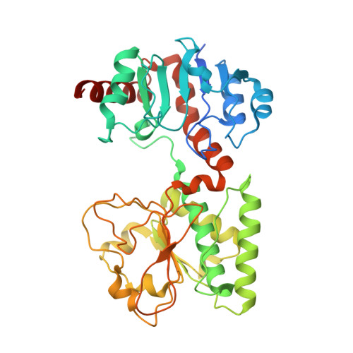
    Three structures of saccharopine dehydrogenase (l-lysine-forming) (SDH) have been determined in the presence of sulfate, adenosine monophosphate (AMP), and oxalylglycine (OxGly). In the sulfate-bound structure, a sulfate ion binds in a cleft between the two domains of SDH, occupies one of the substrate carboxylate binding sites, and results in partial closure of the active site of the enzyme due to a domain rotation of almost 12 degrees in comparison to the apoenzyme structure. In the second structure, AMP binds to the active site in an area where the NAD+ cofactor is expected to bind. All of the AMP moieties (adenine ring, ribose, and phosphate) interact with specific residues of the enzyme. In the OxGly-bound structure, carboxylates of OxGly interact with arginine residues representative of the manner in which substrate (alpha-ketoglutarate and saccharopine) may bind. The alpha-keto group of OxGly interacts with Lys77 and His96, which are candidates for acid-base catalysis. Analysis of ligand-enzyme interactions, comparative structural analysis, corroboration with kinetic data, and discussion of a ternary complex model are presented in this study.


  • Organizational Affiliation
    • Department of Chemistry and Biochemistry, University of Oklahoma, 620 Parrington Oval, Norman, Oklahoma 73019, USA.

Macromolecule Content 

  • Total Structure Weight: 44.4 kDa 
  • Atom Count: 3,245 
  • Modeled Residue Count: 365 
  • Deposited Residue Count: 394 
  • Unique protein chains: 1

Macromolecules

Find similar proteins by:|  3D Structure
Entity ID: 1
MoleculeChains  Sequence LengthOrganismDetailsImage
Saccharopine dehydrogenase [NAD+, L-lysine-forming394Saccharomyces cerevisiaeMutation(s): 0 
Gene Names: LYS1
EC: 1.5.1.7
UniProt
Find proteins for P38998 (Saccharomyces cerevisiae (strain ATCC 204508 / S288c))
Explore P38998 
Go to UniProtKB:  P38998
Entity Groups
Sequence Clusters30% Identity50% Identity70% Identity90% Identity95% Identity100% Identity
UniProt GroupP38998
Sequence Annotations
Expand
Reference Sequence

Small Molecules

Ligands 1 Unique
IDChains Name / Formula / InChI Key2D Diagram3D Interactions
AMP

Query on AMP


Download:Ideal Coordinates CCD File
B [auth A]ADENOSINE MONOPHOSPHATE
C10 H14 N5 O7 P
UDMBCSSLTHHNCD-KQYNXXCUSA-N

Experimental Data & Validation

Experimental Data

  • Method: X-RAY DIFFRACTION
  • Resolution: 1.75 Å
  • R-Value Free:  0.233 (Depositor), 0.231 (DCC) 
  • R-Value Work:  0.199 (Depositor), 0.196 (DCC) 
  • R-Value Observed: 0.200 (Depositor) 
Space Group: P 21 21 21
Unit Cell:
Length ( Å )Angle ( ˚ )
a = 45.97α = 90
b = 69.106β = 90
c = 127.829γ = 90
Software Package:
Software NamePurpose
d*TREKdata scaling
PHASERphasing
REFMACrefinement
PDB_EXTRACTdata extraction
CrystalCleardata collection
d*TREKdata reduction
ARP/wARPmodel building

Structure Validation

View Full Validation Report


Ligand Structure Quality Assessment 


Entry History 

Deposition Data

Revision History  (Full details and data files)

  • Version 1.0: 2007-10-30
    Type: Initial release
  • Version 1.1: 2011-07-13
    Changes: Version format compliance
  • Version 1.2: 2017-10-25
    Changes: Refinement description
  • Version 1.3: 2023-08-30
    Changes: Data collection, Database references, Derived calculations, Refinement description