2QR2

HUMAN QUINONE REDUCTASE TYPE 2, COMPLEX WITH MENADIONE


Experimental Data Snapshot

  • Method: X-RAY DIFFRACTION
  • Resolution: 2.45 Å
  • R-Value Free: 0.274 
  • R-Value Work: 0.218 

wwPDB Validation   3D Report Full Report


This is version 1.3 of the entry. See complete history


Literature

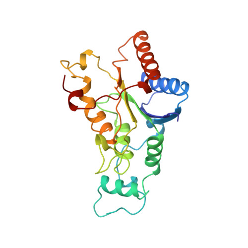
Crystal structure of human quinone reductase type 2, a metalloflavoprotein.

Foster, C.E.Bianchet, M.A.Talalay, P.Zhao, Q.Amzel, L.M.

(1999) Biochemistry 38: 9881-9886

  • DOI: 10.1021/bi990799v
  • Primary Citation of Related Structures:  
    1QR2, 2QR2

  • PubMed Abstract: 
  • In mammals, two separate but homologous cytosolic quinone reductases have been identified: NAD(P)H:quinone oxidoreductase type 1 (QR1) (EC 1.6.99.2) and quinone reductase type 2 (QR2). Although QR1 and QR2 are nearly 50% identical in protein sequence, they display markedly different catalytic properties and substrate specificities ...

    In mammals, two separate but homologous cytosolic quinone reductases have been identified: NAD(P)H:quinone oxidoreductase type 1 (QR1) (EC 1.6.99.2) and quinone reductase type 2 (QR2). Although QR1 and QR2 are nearly 50% identical in protein sequence, they display markedly different catalytic properties and substrate specificities. We report here two crystal structures of QR2: in its native form and bound to menadione (vitamin K(3)), a physiological substrate. Phases were obtained by molecular replacement, using our previously determined rat QR1 structure as the search model. QR2 shares the overall fold of the major catalytic domain of QR1, but lacks the smaller C-terminal domain. The FAD binding sites of QR1 and QR2 are very similar, but their hydride donor binding sites are considerably different. Unexpectedly, we found that QR2 contains a specific metal binding site, which is not present in QR1. Two histidine nitrogens, one cysteine thiol, and a main chain carbonyl group are involved in metal coordination. The metal binding site is solvent-accessible, and is separated from the FAD cofactor by a distance of about 13 A.


    Related Citations: 
    • Unexpected genetic and structural relationships of a long-forgotten flavoenzyme to NAD(P)H:quinone reductase (DT-diaphorase)
      Zhao, Q., Yang, X.L., Holtzclaw, W.D., Talalay, P.
      (1997) Proc Natl Acad Sci U S A 94: 1669

    Organizational Affiliation

    Department of Biophysics, The Johns Hopkins University School of Medicine, Baltimore, Maryland 21205, USA.


Macromolecules
Find similar proteins by:  (by identity cutoff)  |  3D Structure
Entity ID: 1
MoleculeChainsSequence LengthOrganismDetailsImage
PROTEIN (QUINONE REDUCTASE TYPE 2)A, B230Homo sapiensMutation(s): 0 
Gene Names: NQO2NMOR2
EC: 1.6.99.2 (PDB Primary Data), 1.10.5.1 (UniProt)
UniProt & NIH Common Fund Data Resources
Find proteins for P16083 (Homo sapiens)
Explore P16083 
Go to UniProtKB:  P16083
PHAROS:  P16083
Entity Groups  
Sequence Clusters30% Identity50% Identity70% Identity90% Identity95% Identity100% Identity
UniProt GroupP16083
Protein Feature View
Expand
  • Reference Sequence
Small Molecules
Ligands 3 Unique
IDChainsName / Formula / InChI Key2D Diagram3D Interactions
FAD
Query on FAD

Download Ideal Coordinates CCD File 
D [auth A],
F [auth B]
FLAVIN-ADENINE DINUCLEOTIDE
C27 H33 N9 O15 P2
VWWQXMAJTJZDQX-UYBVJOGSSA-N
 Ligand Interaction
VK3
Query on VK3

Download Ideal Coordinates CCD File 
G [auth B],
H [auth B]
MENADIONE
C11 H8 O2
MJVAVZPDRWSRRC-UHFFFAOYSA-N
 Ligand Interaction
ZN
Query on ZN

Download Ideal Coordinates CCD File 
C [auth A],
E [auth B]
ZINC ION
Zn
PTFCDOFLOPIGGS-UHFFFAOYSA-N
 Ligand Interaction
Experimental Data & Validation

Experimental Data

  • Method: X-RAY DIFFRACTION
  • Resolution: 2.45 Å
  • R-Value Free: 0.274 
  • R-Value Work: 0.218 
  • Space Group: P 21 21 21
Unit Cell:
Length ( Å )Angle ( ˚ )
a = 83.6α = 90
b = 106.27β = 90
c = 56.08γ = 90
Software Package:
Software NamePurpose
X-PLORrefinement
DENZOdata reduction
SCALEPACKdata scaling

Structure Validation

View Full Validation Report


Entry History 

Deposition Data

Revision History  (Full details and data files)

  • Version 1.0: 1999-08-18
    Type: Initial release
  • Version 1.1: 2007-10-16
    Changes: Version format compliance
  • Version 1.2: 2011-07-13
    Changes: Derived calculations, Version format compliance
  • Version 1.3: 2019-11-27
    Changes: Database references