


The crystal structure of the human RAC3 in complex with the CRIB domain of human p21-activated kinase 1 (PAK1).
Ugochukwu, E., Yang, X., Elkins, J.M., Burgess-Brown, N., Bunkoczi, G., Knapp, S., Doyle, D.To be published.
Experimental Data Snapshot
Starting Model: experimental
View more details
Entity ID: 1 | |||||
|---|---|---|---|---|---|
| Molecule | Chains | Sequence Length | Organism | Details | Image |
| Ras-related C3 botulinum toxin substrate 3 | 179 | Homo sapiens | Mutation(s): 0 Gene Names: RAC3 EC: 3.6.5.2 |  | |
UniProt & NIH Common Fund Data Resources | |||||
PHAROS: P60763 GTEx: ENSG00000169750 | |||||
Entity Groups | |||||
| Sequence Clusters | 30% Identity50% Identity70% Identity90% Identity95% Identity100% Identity | ||||
| UniProt Group | P60763 | ||||
Sequence AnnotationsExpand | |||||
Reference Sequence | |||||
Entity ID: 2 | |||||
|---|---|---|---|---|---|
| Molecule | Chains | Sequence Length | Organism | Details | Image |
| CRIB domain of the Serine/threonine-protein kinase PAK 1 | B [auth I] | 36 | N/A | Mutation(s): 0 EC: 2.7.11.1 | 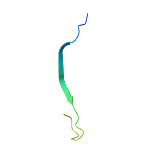 |
UniProt & NIH Common Fund Data Resources | |||||
PHAROS: Q13153 GTEx: ENSG00000149269 | |||||
Entity Groups | |||||
| Sequence Clusters | 30% Identity50% Identity70% Identity90% Identity95% Identity100% Identity | ||||
| UniProt Group | Q13153 | ||||
Sequence AnnotationsExpand | |||||
Reference Sequence | |||||
| Ligands 3 Unique | |||||
|---|---|---|---|---|---|
| ID | Chains | Name / Formula / InChI Key | 2D Diagram | 3D Interactions | |
| GCP Download:Ideal Coordinates CCD File | D [auth A] | PHOSPHOMETHYLPHOSPHONIC ACID GUANYLATE ESTER C11 H18 N5 O13 P3 PHBDHXOBFUBCJD-KQYNXXCUSA-N |  | ||
| GOL Download:Ideal Coordinates CCD File | E [auth A], F [auth A] | GLYCEROL C3 H8 O3 PEDCQBHIVMGVHV-UHFFFAOYSA-N |  | ||
| MG Download:Ideal Coordinates CCD File | C [auth A] | MAGNESIUM ION Mg JLVVSXFLKOJNIY-UHFFFAOYSA-N |  | ||
| Length ( Å ) | Angle ( ˚ ) |
|---|---|
| a = 84.226 | α = 90 |
| b = 53.48 | β = 93.45 |
| c = 47.682 | γ = 90 |
| Software Name | Purpose |
|---|---|
| REFMAC | refinement |
| CrystalClear | data collection |
| MOSFLM | data reduction |
| SCALA | data scaling |
| PHASER | phasing |


