2QKY | pdb_00002qky

complex structure of dipeptidyl peptidase IV and a oxadiazolyl ketone


Experimental Data Snapshot

  • Method: X-RAY DIFFRACTION
  • Resolution: 3.10 Å
  • R-Value Free: 
    0.293 (Depositor) 
  • R-Value Work: 
    0.214 (Depositor) 
  • R-Value Observed: 
    0.214 (Depositor) 

Starting Model: experimental
View more details

wwPDB Validation 3D Report Full Report

Validation slider image for 2QKY

This is version 1.4 of the entry. See complete history

Literature

Synthesis, SAR, and X-ray structure of novel potent DPPIV inhibitors: oxadiazolyl ketones.

Koo, K.D.Kim, M.J.Kim, S.Kim, K.H.Hong, S.Y.Hur, G.C.Yim, H.J.Kim, G.T.Han, H.O.Kwon, O.H.Kwon, T.S.Koh, J.S.Lee, C.S.

(2007) Bioorg Med Chem Lett 17: 4167-4172

  • DOI: https://doi.org/10.1016/j.bmcl.2007.05.046
  • Primary Citation Related Structures: 
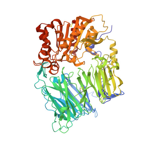
    2QKY

  • PubMed Abstract: 

    Synthesis of a novel series of DPPIV inhibitors with 1,2,4- and 1,3,4-oxadiazolyl ketone derivatives and its structure-activity relationships are discussed. Compound 18h showed good inhibitory activity against DPPIV and favorable pharmacokinetic properties. In vivo pharmacodynamic efficacy and co-crystal structure of compound 18h with DPPIV is also described.


  • Organizational Affiliation
    • LG Life Sciences, Ltd/R&D Park, 104-1 Munji-dong, Yuseong-gu, Daejeon 305-380, Republic of Korea.

Macromolecule Content 

  • Total Structure Weight: 339.4 kDa 
  • Atom Count: 24,275 
  • Modeled Residue Count: 2,912 
  • Deposited Residue Count: 2,912 
  • Unique protein chains: 1

Macromolecules

Find similar proteins by:|  3D Structure
Entity ID: 1
MoleculeChains  Sequence LengthOrganismDetailsImage
Dipeptidyl peptidase 4 (EC 3.4.14.5) (Dipeptidyl peptidase IV) (DPP IV) (T-cell activation antigen CD26) (TP103) (Adenosine deaminase complexing protein 2) (ADABP) (Dipeptidyl peptidase 4 soluble form) (Dipeptidyl peptidase IV soluble form)
A, B, C, D
728Homo sapiensMutation(s): 0 
Gene Names: DPP4ADCP2CD26
EC: 3.4.14.5
UniProt & NIH Common Fund Data Resources
Find proteins for P27487 (Homo sapiens)
Explore P27487 
Go to UniProtKB:  P27487
PHAROS:  P27487
GTEx:  ENSG00000197635 
Entity Groups
Sequence Clusters30% Identity50% Identity70% Identity90% Identity95% Identity100% Identity
UniProt GroupP27487
Sequence Annotations
Expand
Reference Sequence

Small Molecules

Ligands 1 Unique
IDChains Name / Formula / InChI Key2D Diagram3D Interactions
13Z

Query on 13Z


Download:Ideal Coordinates CCD File
E [auth A],
F [auth B],
G [auth C],
H [auth D]
2-[(2-{(2S,4S)-2-[(R)-(5-tert-butyl-1,3,4-oxadiazol-2-yl)(hydroxy)methyl]-4-fluoropyrrolidin-1-yl}-2-oxoethyl)amino]-2-methylpropan-1-ol
C17 H29 F N4 O4
PHTMASAWZAGGEK-GMXVVIOVSA-N

Experimental Data & Validation

Experimental Data

  • Method: X-RAY DIFFRACTION
  • Resolution: 3.10 Å
  • R-Value Free:  0.293 (Depositor) 
  • R-Value Work:  0.214 (Depositor) 
  • R-Value Observed: 0.214 (Depositor) 
Space Group: P 1
Unit Cell:
Length ( Å )Angle ( ˚ )
a = 73.178α = 76.3
b = 106.115β = 78.62
c = 132.051γ = 80.11
Software Package:
Software NamePurpose
DENZOdata reduction
SCALEPACKdata scaling
CNSrefinement
PDB_EXTRACTdata extraction
HKL-2000data collection
HKL-2000data reduction
AMoREphasing

Structure Validation

View Full Validation Report


Entry History 

Deposition Data

Revision History  (Full details and data files)

  • Version 1.0: 2008-07-15
    Type: Initial release
  • Version 1.1: 2011-07-13
    Changes: Version format compliance
  • Version 1.2: 2017-10-18
    Changes: Refinement description
  • Version 1.3: 2023-08-30
    Changes: Data collection, Database references, Derived calculations, Refinement description
  • Version 1.4: 2024-11-20
    Changes: Structure summary