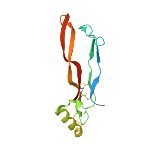
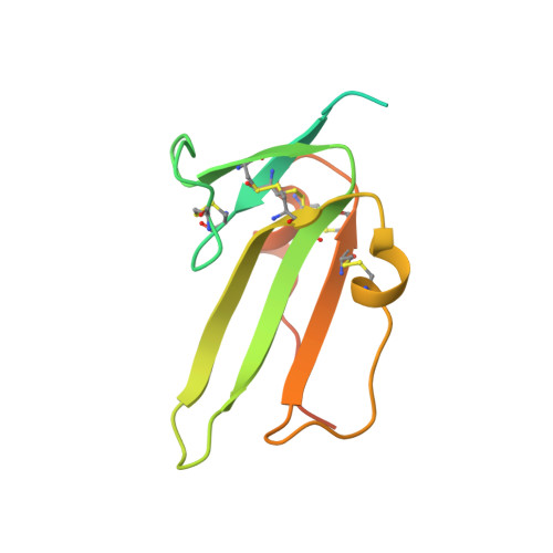
2QJB

Crystal structure analysis of BMP-2 in complex with BMPR-IA variant IA/IB


Experimental Data Snapshot

  • Method: X-RAY DIFFRACTION
  • Resolution: 2.50 Å
  • R-Value Free: 0.263 
  • R-Value Work: 0.204 
  • R-Value Observed: 0.207 

Starting Model: experimental
View more details

wwPDB Validation   3D Report Full Report


This is version 1.4 of the entry. See complete history


Literature

Structure analysis of bone morphogenetic protein-2 type I receptor complexes reveals a mechanism of receptor inactivation in juvenile polyposis syndrome.

Kotzsch, A.Nickel, J.Seher, A.Heinecke, K.van Geersdaele, L.Herrmann, T.Sebald, W.Mueller, T.D.

(2008) J Biol Chem 283: 5876-5887

  • DOI: https://doi.org/10.1074/jbc.M706029200
  • Primary Citation of Related Structures:  
    2QJ9, 2QJA, 2QJB

  • PubMed Abstract: 

    Bone morphogenetic proteins regulate many developmental processes during embryogenesis as well as tissue homeostasis in the adult. Signaling of bone morphogenetic proteins (BMPs) is accomplished by binding to two types of serine/threonine kinase transmembrane receptors termed type I and type II. Because a large number of ligands signal through a limited number of receptors, ligand-receptor interaction in the BMP superfamily is highly promiscuous, with a ligand binding to various receptors and a receptor binding many different BMP ligands. In this study we investigate the interaction of BMP-2 with its two high affinity type I receptors, BMP receptors IA (BMPR-IA) and BMPR-IB. Interestingly, 50% of the residues in the BMP-2 binding epitope of the BMPR-IA receptor are exchanged in BMPR-IB without a decrease in binding affinity or specificity for BMP-2. Our structural and functional analyses show that promiscuous binding of BMP-2 to both type I receptors is achieved by inherent backbone and side-chain flexibility as well as by variable hydration of the ligand-receptor interface enabling the BMP-2 surface to adapt to different receptor geometries. Despite the high degree of amino acid variability found in BMPR-IA and BMPR-IB binding equally to BMP-2, three single point missense mutations in the ectodomain of BMPR-IA cannot be tolerated. In juvenile polyposis syndrome these mutations have been shown to inactivate BMPR-IA. On the basis of our biochemical and biophysical analyses, we can show that the mutations, which are located outside the ligand binding epitope, alter the local or global fold of the receptor, thereby inactivating BMPR-IA and causing a loss of the BMP-2 tumor suppressor function in colon epithelial cells.


  • Organizational Affiliation

    Lehrstuhl für Botanik I-Molekulare Pflanzenphysiologie und Biophysik, Julius-von-Sachs-Institut der Universität Würzburg, Julius-von-Sachs-Platz 2, D-97082 Würzburg, Germany.


Macromolecules
Find similar proteins by:  (by identity cutoff)  |  3D Structure
Entity ID: 1
MoleculeChains Sequence LengthOrganismDetailsImage
Bone morphogenetic protein 2
A, B
116Homo sapiensMutation(s): 0 
Gene Names: BMP2BMP2A
UniProt & NIH Common Fund Data Resources
Find proteins for P12643 (Homo sapiens)
Explore P12643 
Go to UniProtKB:  P12643
PHAROS:  P12643
GTEx:  ENSG00000125845 
Entity Groups  
Sequence Clusters30% Identity50% Identity70% Identity90% Identity95% Identity100% Identity
UniProt GroupP12643
Sequence Annotations
Expand
  • Reference Sequence
Find similar proteins by:  (by identity cutoff)  |  3D Structure
Entity ID: 2
MoleculeChains Sequence LengthOrganismDetailsImage
Bone morphogenetic protein receptor type IA
C, D
135Homo sapiensMutation(s): 15 
Gene Names: BMPR1AACVRLK3ALK3
EC: 2.7.11.30
UniProt & NIH Common Fund Data Resources
Find proteins for P36894 (Homo sapiens)
Explore P36894 
Go to UniProtKB:  P36894
PHAROS:  P36894
GTEx:  ENSG00000107779 
Entity Groups  
Sequence Clusters30% Identity50% Identity70% Identity90% Identity95% Identity100% Identity
UniProt GroupP36894
Sequence Annotations
Expand
  • Reference Sequence
Small Molecules
Ligands 1 Unique
IDChains Name / Formula / InChI Key2D Diagram3D Interactions
CL
Query on CL

Download Ideal Coordinates CCD File 
E [auth B]CHLORIDE ION
Cl
VEXZGXHMUGYJMC-UHFFFAOYSA-M
Experimental Data & Validation

Experimental Data

  • Method: X-RAY DIFFRACTION
  • Resolution: 2.50 Å
  • R-Value Free: 0.263 
  • R-Value Work: 0.204 
  • R-Value Observed: 0.207 
  • Space Group: P 65
Unit Cell:
Length ( Å )Angle ( ˚ )
a = 104.175α = 90
b = 104.175β = 90
c = 100.012γ = 120
Software Package:
Software NamePurpose
REFMACrefinement
CrystalCleardata collection
CrystalCleardata reduction
CrystalCleardata scaling
PHASERphasing

Structure Validation

View Full Validation Report


Entry History 

Deposition Data

Revision History  (Full details and data files)

  • Version 1.0: 2008-01-15
    Type: Initial release
  • Version 1.1: 2011-07-13
    Changes: Advisory, Version format compliance
  • Version 1.2: 2021-10-20
    Changes: Database references
  • Version 1.3: 2023-08-30
    Changes: Data collection, Refinement description
  • Version 1.4: 2024-11-13
    Changes: Structure summary