2QIH

Crystal structure of 527-665 fragment of UspA1 protein from Moraxella catarrhalis


Experimental Data Snapshot

  • Method: X-RAY DIFFRACTION
  • Resolution: 1.90 Å
  • R-Value Free: 0.253 
  • R-Value Work: 0.194 
  • R-Value Observed: 0.197 

wwPDB Validation   3D Report Full Report


This is version 1.4 of the entry. See complete history


Literature

The Moraxella adhesin UspA1 binds to its human CEACAM1 receptor by a deformable trimeric coiled-coil.

Conners, R.Hill, D.J.Borodina, E.Agnew, C.Daniell, S.J.Burton, N.M.Sessions, R.B.Clarke, A.R.Catto, L.E.Lammie, D.Wess, T.Brady, R.L.Virji, M.

(2008) EMBO J 27: 1779-1789

  • DOI: https://doi.org/10.1038/emboj.2008.101
  • Primary Citation of Related Structures:  
    2QIH

  • PubMed Abstract: 

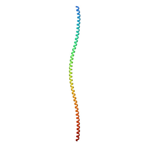
    Moraxella catarrhalis is a ubiquitous human-specific bacterium commonly associated with upper and lower respiratory tract infections, including otitis media, sinusitis and chronic obstructive pulmonary disease. The bacterium uses an autotransporter protein UspA1 to target an important human cellular receptor carcinoembryonic antigen-related cell adhesion molecule 1 (CEACAM1). Using X-ray crystallography, we show that the CEACAM1 receptor-binding region of UspA1 unusually consists of an extended, rod-like left-handed trimeric coiled-coil. Mutagenesis and binding studies of UspA1 and the N-domain of CEACAM1 have been used to delineate the interacting surfaces between ligand and receptor and guide assembly of the complex. However, solution scattering, molecular modelling and electron microscopy analyses all indicate that significant bending of the UspA1 coiled-coil stalk also occurs. This explains how UspA1 can engage CEACAM1 at a site far distant from its head group, permitting closer proximity of the respective cell surfaces during infection.


  • Organizational Affiliation

    Department of Biochemistry, University of Bristol, Bristol, UK.


Macromolecules
Find similar proteins by:  (by identity cutoff)  |  3D Structure
Entity ID: 1
MoleculeChains Sequence LengthOrganismDetailsImage
protein UspA1
A, B
157Moraxella catarrhalisMutation(s): 1 
Gene Names: UspA1
UniProt
Find proteins for Q9XD54 (Moraxella catarrhalis)
Explore Q9XD54 
Go to UniProtKB:  Q9XD54
Entity Groups  
Sequence Clusters30% Identity50% Identity70% Identity90% Identity95% Identity100% Identity
UniProt GroupQ9XD54
Sequence Annotations
Expand
  • Reference Sequence
Small Molecules
Ligands 2 Unique
IDChains Name / Formula / InChI Key2D Diagram3D Interactions
PO4
Query on PO4

Download Ideal Coordinates CCD File 
C [auth A],
D [auth A],
M [auth B],
N [auth B]
PHOSPHATE ION
O4 P
NBIIXXVUZAFLBC-UHFFFAOYSA-K
CL
Query on CL

Download Ideal Coordinates CCD File 
E [auth A]
F [auth A]
G [auth A]
H [auth A]
I [auth A]
E [auth A],
F [auth A],
G [auth A],
H [auth A],
I [auth A],
J [auth A],
K [auth A],
L [auth A],
O [auth B],
P [auth B],
Q [auth B],
R [auth B],
S [auth B],
T [auth B],
U [auth B],
V [auth B]
CHLORIDE ION
Cl
VEXZGXHMUGYJMC-UHFFFAOYSA-M
Experimental Data & Validation

Experimental Data

  • Method: X-RAY DIFFRACTION
  • Resolution: 1.90 Å
  • R-Value Free: 0.253 
  • R-Value Work: 0.194 
  • R-Value Observed: 0.197 
  • Space Group: P 3
Unit Cell:
Length ( Å )Angle ( ˚ )
a = 37.234α = 90
b = 37.234β = 90
c = 224.475γ = 120
Software Package:
Software NamePurpose
DENZOdata reduction
SCALEPACKdata scaling
PHASERphasing
REFMACrefinement
PDB_EXTRACTdata extraction
HKL-2000data collection
HKL-2000data reduction

Structure Validation

View Full Validation Report


Entry History 

Deposition Data

Revision History  (Full details and data files)

  • Version 1.0: 2008-05-20
    Type: Initial release
  • Version 1.1: 2011-07-13
    Changes: Version format compliance
  • Version 1.2: 2017-10-18
    Changes: Refinement description
  • Version 1.3: 2021-10-20
    Changes: Database references, Derived calculations
  • Version 1.4: 2023-08-30
    Changes: Data collection, Refinement description