2QFI | pdb_00002qfi

Structure of the zinc transporter YiiP


Experimental Data Snapshot

  • Method: X-RAY DIFFRACTION
  • Resolution: 3.80 Å
  • R-Value Free: 
    0.329 (Depositor), 0.333 (DCC) 
  • R-Value Work: 
    0.322 (Depositor), 0.348 (DCC) 
  • R-Value Observed: 
    0.323 (Depositor) 

wwPDB Validation 3D Report Full Report

Validation slider image for 2QFI

This is version 1.2 of the entry. See complete history

Literature

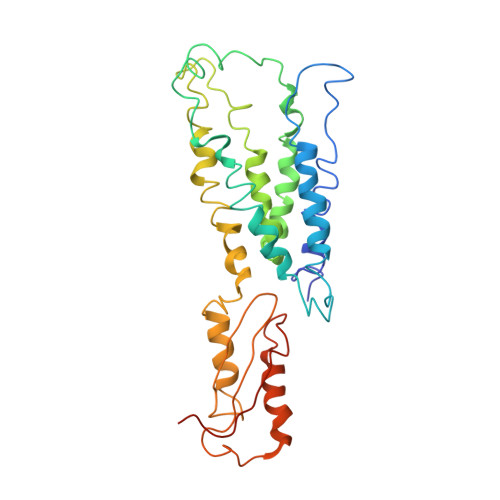
Structure of the zinc transporter YiiP.

Lu, M.Fu, D.

(2007) Science 317: 1746-1748

  • DOI: https://doi.org/10.1126/science.1143748
  • Primary Citation Related Structures: 
    2QFI

  • PubMed Abstract: 

    YiiP is a membrane transporter that catalyzes Zn2+/H+ exchange across the inner membrane of Escherichia coli. Mammalian homologs of YiiP play critical roles in zinc homeostasis and cell signaling. Here, we report the x-ray structure of YiiP in complex with zinc at 3.8 angstrom resolution. YiiP is a homodimer held together in a parallel orientation through four Zn2+ ions at the interface of the cytoplasmic domains, whereas the two transmembrane domains swing out to yield a Y-shaped structure. In each protomer, the cytoplasmic domain adopts a metallochaperone-like protein fold; the transmembrane domain features a bundle of six transmembrane helices and a tetrahedral Zn2+ binding site located in a cavity that is open to both the membrane outer leaflet and the periplasm.


  • Organizational Affiliation
    • Department of Biology, Brookhaven National Laboratory, Upton, NY 11973, USA.

Macromolecule Content 

  • Total Structure Weight: 66.37 kDa 
  • Atom Count: 4,417 
  • Modeled Residue Count: 572 
  • Deposited Residue Count: 600 
  • Unique protein chains: 1

Macromolecules

Find similar proteins by:|  3D Structure
Entity ID: 1
MoleculeChains  Sequence LengthOrganismDetailsImage
Ferrous-iron efflux pump fieF
A, B
300Escherichia coliMutation(s): 0 
Gene Names: fieF
Membrane Entity: Yes 
UniProt
Find proteins for P69380 (Escherichia coli (strain K12))
Explore P69380 
Go to UniProtKB:  P69380
Entity Groups
Sequence Clusters30% Identity50% Identity70% Identity90% Identity95% Identity100% Identity
UniProt GroupP69380
Sequence Annotations
Expand
Reference Sequence

Experimental Data & Validation

Experimental Data

  • Method: X-RAY DIFFRACTION
  • Resolution: 3.80 Å
  • R-Value Free:  0.329 (Depositor), 0.333 (DCC) 
  • R-Value Work:  0.322 (Depositor), 0.348 (DCC) 
  • R-Value Observed: 0.323 (Depositor) 
Space Group: P 2 2 21
Unit Cell:
Length ( Å )Angle ( ˚ )
a = 106.66α = 90
b = 110.839β = 90
c = 130.662γ = 90
Software Package:
Software NamePurpose
REFMACrefinement
HKL-2000data collection
HKL-2000data reduction
SCALAdata scaling
SHARPphasing

Structure Validation

View Full Validation Report


Entry History 

Deposition Data

  • Released Date: 2007-10-16 
  • Deposition Author(s): Lu, M.

Revision History  (Full details and data files)

  • Version 1.0: 2007-10-16
    Type: Initial release
  • Version 1.1: 2011-07-13
    Changes: Derived calculations, Version format compliance
  • Version 1.2: 2024-02-21
    Changes: Data collection, Database references, Derived calculations, Refinement description