2QBY | pdb_00002qby

Crystal structure of a heterodimer of Cdc6/Orc1 initiators bound to origin DNA (from S. solfataricus)


Experimental Data Snapshot

  • Method: X-RAY DIFFRACTION
  • Resolution: 3.35 Å
  • R-Value Free: 
    0.269 (Depositor), 0.262 (DCC) 
  • R-Value Work: 
    0.226 (Depositor), 0.220 (DCC) 
  • R-Value Observed: 
    0.226 (Depositor) 

Starting Model: experimental
View more details

wwPDB Validation 3D Report Full Report

Validation slider image for 2QBY

Ligand Structure Quality Assessment 


This is version 1.2 of the entry. See complete history

Literature

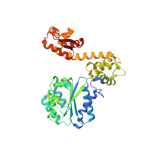
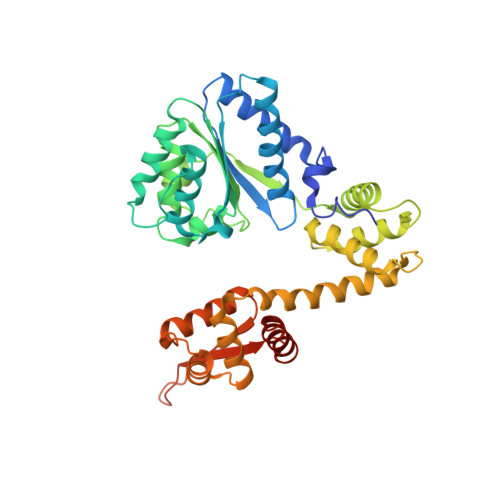
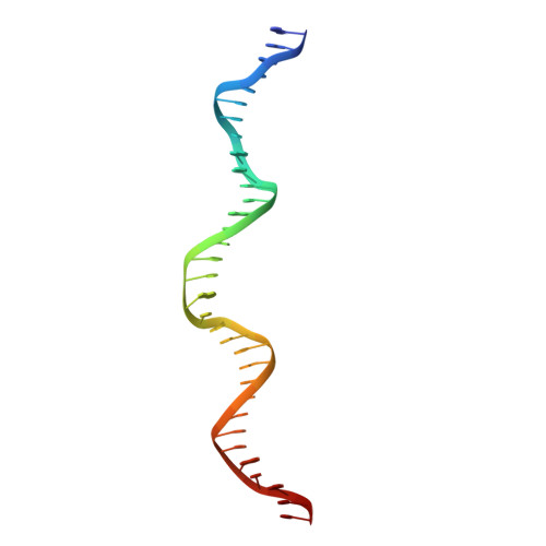
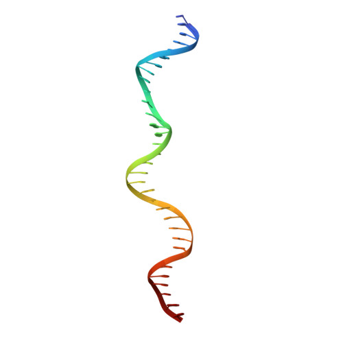
Replication origin recognition and deformation by a heterodimeric archaeal Orc1 complex.

Dueber, E.L.Corn, J.E.Bell, S.D.Berger, J.M.

(2007) Science 317: 1210-1213

  • DOI: https://doi.org/10.1126/science.1143690
  • Primary Citation Related Structures: 
    2QBY

  • PubMed Abstract: 

    The faithful duplication of genetic material depends on essential DNA replication initiation factors. Cellular initiators form higher-order assemblies on replication origins, using adenosine triphosphate (ATP) to locally remodel duplex DNA and facilitate proper loading of synthetic replisomal components. To better understand initiator function, we determined the 3.4 angstrom-resolution structure of an archaeal Cdc6/Orc1 heterodimer bound to origin DNA. The structure demonstrates that, in addition to conventional DNA binding elements, initiators use their AAA+ ATPase domains to recognize origin DNA. Together these interactions establish the polarity of initiator assembly on the origin and induce substantial distortions into origin DNA strands. Biochemical and comparative analyses indicate that AAA+/DNA contacts observed in the structure are dynamic and evolutionarily conserved, suggesting that the complex forms a core component of the basal initiation machinery.


  • Organizational Affiliation
    • Miller Institute for Basic Research in Science, 2536 Channing Way 5190, University of California, Berkeley, CA 94720, USA.

Macromolecule Content 

  • Total Structure Weight: 108.92 kDa 
  • Atom Count: 7,309 
  • Modeled Residue Count: 800 
  • Deposited Residue Count: 836 
  • Unique protein chains: 2
  • Unique nucleic acid chains: 2

Macromolecules


Find similar proteins by:|  3D Structure
Entity ID: 3
MoleculeChains  Sequence LengthOrganismDetailsImage
Cell division control protein 6 homolog 1C [auth A]386Saccharolobus solfataricusMutation(s): 0 
Gene Names: cdc6-1
UniProt
Find proteins for Q980N4 (Saccharolobus solfataricus (strain ATCC 35092 / DSM 1617 / JCM 11322 / P2))
Explore Q980N4 
Go to UniProtKB:  Q980N4
Entity Groups
Sequence Clusters30% Identity50% Identity70% Identity90% Identity95% Identity100% Identity
UniProt GroupQ980N4
Sequence Annotations
Expand
Reference Sequence
Find similar proteins by:|  3D Structure
Entity ID: 4
MoleculeChains  Sequence LengthOrganismDetailsImage
Cell division control protein 6 homolog 3D [auth B]384Saccharolobus solfataricusMutation(s): 0 
Gene Names: cdc6-3
UniProt
Find proteins for Q97WM8 (Saccharolobus solfataricus (strain ATCC 35092 / DSM 1617 / JCM 11322 / P2))
Explore Q97WM8 
Go to UniProtKB:  Q97WM8
Entity Groups
Sequence Clusters30% Identity50% Identity70% Identity90% Identity95% Identity100% Identity
UniProt GroupQ97WM8
Sequence Annotations
Expand
Reference Sequence
Find similar nucleic acids by:  (by identity cutoff) 
Entity ID: 1
MoleculeChains LengthOrganismImage
DNA (33-MER)A [auth C]33N/A
Sequence Annotations
Expand
Reference Sequence
Find similar nucleic acids by:  (by identity cutoff) 
Entity ID: 2
MoleculeChains LengthOrganismImage
DNA (33-MER)B [auth D]33N/A
Sequence Annotations
Expand
Reference Sequence

Small Molecules

Ligands 4 Unique
IDChains Name / Formula / InChI Key2D Diagram3D Interactions
ADP

Query on ADP


Download:Ideal Coordinates CCD File
I [auth A],
K [auth B]
ADENOSINE-5'-DIPHOSPHATE
C10 H15 N5 O10 P2
XTWYTFMLZFPYCI-KQYNXXCUSA-N
SPD

Query on SPD


Download:Ideal Coordinates CCD File
E [auth C],
F [auth C]
SPERMIDINE
C7 H19 N3
ATHGHQPFGPMSJY-UHFFFAOYSA-N
K

Query on K


Download:Ideal Coordinates CCD File
G [auth D]POTASSIUM ION
K
NPYPAHLBTDXSSS-UHFFFAOYSA-N
MG

Query on MG


Download:Ideal Coordinates CCD File
H [auth A],
J [auth B]
MAGNESIUM ION
Mg
JLVVSXFLKOJNIY-UHFFFAOYSA-N

Experimental Data & Validation

Experimental Data

  • Method: X-RAY DIFFRACTION
  • Resolution: 3.35 Å
  • R-Value Free:  0.269 (Depositor), 0.262 (DCC) 
  • R-Value Work:  0.226 (Depositor), 0.220 (DCC) 
  • R-Value Observed: 0.226 (Depositor) 
Space Group: C 2 2 21
Unit Cell:
Length ( Å )Angle ( ˚ )
a = 77.649α = 90
b = 199.143β = 90
c = 213.405γ = 90
Software Package:
Software NamePurpose
HKL-2000data collection
MLPHAREphasing
PHENIXrefinement
HKL-2000data reduction
SCALEPACKdata scaling

Structure Validation

View Full Validation Report


Ligand Structure Quality Assessment 


Entry History 

Deposition Data

Revision History  (Full details and data files)

  • Version 1.0: 2007-09-11
    Type: Initial release
  • Version 1.1: 2011-07-13
    Changes: Version format compliance
  • Version 1.2: 2023-08-30
    Changes: Data collection, Database references, Derived calculations, Refinement description