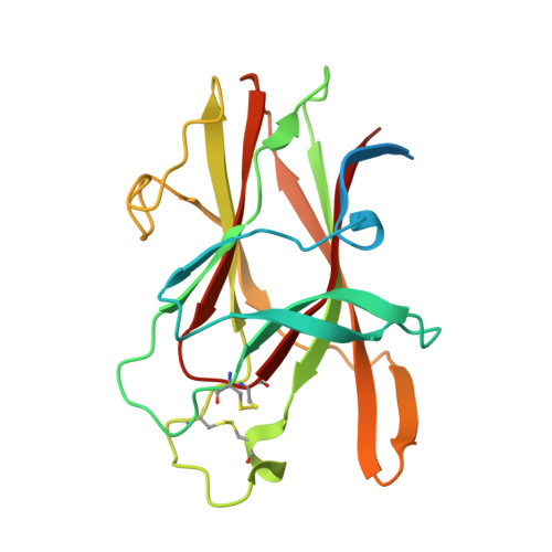
2QBX

EphB2/SNEW Antagonistic Peptide Complex


Experimental Data Snapshot

  • Method: X-RAY DIFFRACTION
  • Resolution: 2.30 Å
  • R-Value Free: 0.270 
  • R-Value Work: 0.194 
  • R-Value Observed: 0.198 

wwPDB Validation   3D Report Full Report


This is version 1.2 of the entry. See complete history


Literature

Three-dimensional structure of the EphB2 receptor in complex with an antagonistic peptide reveals a novel mode of inhibition.

Chrencik, J.E.Brooun, A.Recht, M.I.Nicola, G.Davis, L.K.Abagyan, R.Widmer, H.Pasquale, E.B.Kuhn, P.

(2007) J Biol Chem 282: 36505-36513

  • DOI: https://doi.org/10.1074/jbc.M706340200
  • Primary Citation of Related Structures:  
    2QBX

  • PubMed Abstract: 

    The Eph family of receptor tyrosine kinases has been implicated in tumorigenesis as well as pathological forms of angiogenesis. Understanding how to modulate the interaction of Eph receptors with their ephrin ligands is therefore of critical interest for the development of therapeutics to treat cancer. Previous work identified a set of 12-mer peptides that displayed moderate binding affinity but high selectivity for the EphB2 receptor. The SNEW antagonistic peptide inhibited the interaction of EphB2 with ephrinB2, with an IC50 of approximately 15 microm. To gain a better molecular understanding of how to inhibit Eph/ephrin binding, we determined the crystal structure of the EphB2 receptor in complex with the SNEW peptide to 2.3-A resolution. The peptide binds in the hydrophobic ligand-binding cleft of the EphB2 receptor, thus competing with the ephrin ligand for receptor binding. However, the binding interactions of the SNEW peptide are markedly different from those described for the TNYL-RAW peptide, which binds to the ligand-binding cleft of EphB4, indicating a novel mode of antagonism. Nevertheless, we identified a conserved structural motif present in all known receptor/ligand interfaces, which may serve as a scaffold for the development of therapeutic leads. The EphB2-SNEW complex crystallized as a homodimer, and the residues involved in the dimerization interface are similar to those implicated in mediating tetramerization of EphB2-ephrinB2 complexes. The structure of EphB2 in complex with the SNEW peptide reveals novel binding determinants that could serve as starting points in the development of compounds that modulate Eph receptor/ephrin interactions and biological activities.


  • Organizational Affiliation

    Department of Cellular Biology and Molecular Biology, The Scripps Research Institute, La Jolla, California 92037, USA.


Macromolecules
Find similar proteins by:  (by identity cutoff)  |  3D Structure
Entity ID: 1
MoleculeChains Sequence LengthOrganismDetailsImage
Ephrin type-B receptor 2
A, B
208Homo sapiensMutation(s): 0 
Gene Names: EPHB2DRTEPHT3EPTH3ERKHEK5TYRO5
EC: 2.7.10.1
UniProt & NIH Common Fund Data Resources
Find proteins for P29323 (Homo sapiens)
Explore P29323 
Go to UniProtKB:  P29323
PHAROS:  P29323
GTEx:  ENSG00000133216 
Entity Groups  
Sequence Clusters30% Identity50% Identity70% Identity90% Identity95% Identity100% Identity
UniProt GroupP29323
Sequence Annotations
Expand
  • Reference Sequence

Find similar proteins by:  Sequence   |   3D Structure  

Entity ID: 2
MoleculeChains Sequence LengthOrganismDetailsImage
antagonistic peptideC [auth D],
D [auth P]
12N/AMutation(s): 0 
Entity Groups  
Sequence Clusters30% Identity50% Identity70% Identity90% Identity95% Identity100% Identity
Sequence Annotations
Expand
  • Reference Sequence
Small Molecules
Ligands 1 Unique
IDChains Name / Formula / InChI Key2D Diagram3D Interactions
SO4
Query on SO4

Download Ideal Coordinates CCD File 
E [auth A]
F [auth A]
G [auth A]
H [auth A]
I [auth B]
E [auth A],
F [auth A],
G [auth A],
H [auth A],
I [auth B],
J [auth B],
K [auth B],
L [auth B],
M [auth B],
N [auth B],
O [auth B],
P [auth D]
SULFATE ION
O4 S
QAOWNCQODCNURD-UHFFFAOYSA-L
Experimental Data & Validation

Experimental Data

  • Method: X-RAY DIFFRACTION
  • Resolution: 2.30 Å
  • R-Value Free: 0.270 
  • R-Value Work: 0.194 
  • R-Value Observed: 0.198 
  • Space Group: P 32
Unit Cell:
Length ( Å )Angle ( ˚ )
a = 40.183α = 90
b = 40.183β = 90
c = 235.025γ = 120
Software Package:
Software NamePurpose
REFMACrefinement
HKL-2000data collection
HKL-2000data reduction
HKL-2000data scaling
AMoREphasing

Structure Validation

View Full Validation Report


Entry History 

Revision History  (Full details and data files)

  • Version 1.0: 2007-11-06
    Type: Initial release
  • Version 1.1: 2011-07-13
    Changes: Version format compliance
  • Version 1.2: 2023-08-30
    Changes: Data collection, Database references, Derived calculations, Refinement description