2Q94

E. coli methionine aminopeptidase Mn-form with inhibitor A04


Experimental Data Snapshot

  • Method: X-RAY DIFFRACTION
  • Resolution: 1.63 Å
  • R-Value Free: 0.238 
  • R-Value Work: 0.213 

wwPDB Validation   3D Report Full Report


Ligand Structure Quality Assessment 


This is version 1.2 of the entry. See complete history


Literature

Structural analysis of inhibition of E. coli methionine aminopeptidase: implication of loop flexibility in selective inhibition of bacterial enzymes.

Ma, Z.Q.Xie, S.X.Huang, Q.Q.Nan, F.J.Hurley, T.D.Ye, Q.Z.

(2007) BMC Struct Biol 7: 84-84

  • DOI: https://doi.org/10.1186/1472-6807-7-84
  • Primary Citation of Related Structures:  
    2Q92, 2Q93, 2Q94, 2Q95, 2Q96

  • PubMed Abstract: 

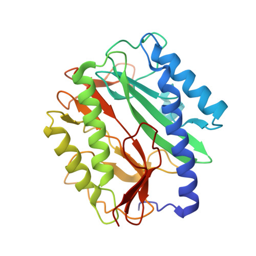
    Methionine aminopeptidase is a potential target of future antibacterial and anticancer drugs. Structural analysis of complexes of the enzyme with its inhibitors provides valuable information for structure-based drug design efforts. Five new X-ray structures of such enzyme-inhibitor complexes were obtained. Analysis of these and other three similar structures reveals the adaptability of a surface-exposed loop bearing Y62, H63, G64 and Y65 (the YHGY loop) that is an integral part of the substrate and inhibitor binding pocket. This adaptability is important for accommodating inhibitors with variations in size. When compared with the human isozymes, this loop either becomes buried in the human type I enzyme due to an N-terminal extension that covers its position or is replaced by a unique insert in the human type II enzyme. The adaptability of the YHGY loop in E. coli methionine aminopeptidase, and likely in other bacterial methionine aminopeptidases, enables the enzyme active pocket to accommodate inhibitors of differing size. The differences in this adaptable loop between the bacterial and human methionine aminopeptidases is a structural feature that can be exploited to design inhibitors of bacterial methionine aminopeptidases as therapeutic agents with minimal inhibition of the corresponding human enzymes.


  • Organizational Affiliation

    High Throughput Screening Laboratory, University of Kansas, Lawrence, Kansas 66047, USA. zeqiang.ma@vanderbilt.edu


Macromolecules
Find similar proteins by:  (by identity cutoff)  |  3D Structure
Entity ID: 1
MoleculeChains Sequence LengthOrganismDetailsImage
Methionine aminopeptidase262Escherichia coliMutation(s): 0 
Gene Names: map
EC: 3.4.11.18
UniProt
Find proteins for P0AE18 (Escherichia coli (strain K12))
Explore P0AE18 
Go to UniProtKB:  P0AE18
Entity Groups  
Sequence Clusters30% Identity50% Identity70% Identity90% Identity95% Identity100% Identity
UniProt GroupP0AE18
Sequence Annotations
Expand
  • Reference Sequence
Small Molecules
Binding Affinity Annotations 
IDSourceBinding Affinity
A04 Binding MOAD:  2Q94 IC50: 370 (nM) from 1 assay(s)
PDBBind:  2Q94 IC50: 370 (nM) from 1 assay(s)
BindingDB:  2Q94 IC50: min: 368, max: 1.97e+5 (nM) from 4 assay(s)
Experimental Data & Validation

Experimental Data

  • Method: X-RAY DIFFRACTION
  • Resolution: 1.63 Å
  • R-Value Free: 0.238 
  • R-Value Work: 0.213 
  • Space Group: P 1 21 1
Unit Cell:
Length ( Å )Angle ( ˚ )
a = 38.199α = 90
b = 60.605β = 104.8
c = 50.624γ = 90
Software Package:
Software NamePurpose
SCALAdata scaling
MOLREPphasing
CNSrefinement
PDB_EXTRACTdata extraction
MOSFLMdata reduction

Structure Validation

View Full Validation Report


Ligand Structure Quality Assessment 


Entry History 

Deposition Data

  • Released Date: 2008-01-01 
  • Deposition Author(s): Ye, Q.-Z.

Revision History  (Full details and data files)

  • Version 1.0: 2008-01-01
    Type: Initial release
  • Version 1.1: 2011-07-13
    Changes: Version format compliance
  • Version 1.2: 2023-08-30
    Changes: Data collection, Database references, Derived calculations, Refinement description