2Q7W | pdb_00002q7w

Structural Studies Reveals the Inactivation of E. coli L-aspartate aminotransferase (S)-4,5-amino-dihydro-2-thiophenecarboxylic acid (SADTA) via two mechanisms at pH 6.0


Experimental Data Snapshot

  • Method: X-RAY DIFFRACTION
  • Resolution: 1.40 Å
  • R-Value Free: 
    0.188 (Depositor), 0.187 (DCC) 
  • R-Value Work: 
    0.154 (Depositor), 0.152 (DCC) 
  • R-Value Observed: 
    0.156 (Depositor) 

Starting Model: experimental
View more details

wwPDB Validation 3D Report Full Report

Validation slider image for 2Q7W

Ligand Structure Quality Assessment 


This is version 1.2 of the entry. See complete history

Literature

Inactivation of Escherichia coli l-Aspartate Aminotransferase by (S)-4-Amino-4,5-dihydro-2-thiophenecarboxylic Acid Reveals "A Tale of Two Mechanisms".

Liu, D.Pozharski, E.Lepore, B.W.Fu, M.Silverman, R.B.Petsko, G.A.Ringe, D.

(2007) Biochemistry 46: 10517-10527

  • DOI: https://doi.org/10.1021/bi700663n
  • Primary Citation Related Structures: 
    2Q7W, 2QA3, 2QB2, 2QB3, 2QBT

  • PubMed Abstract: 

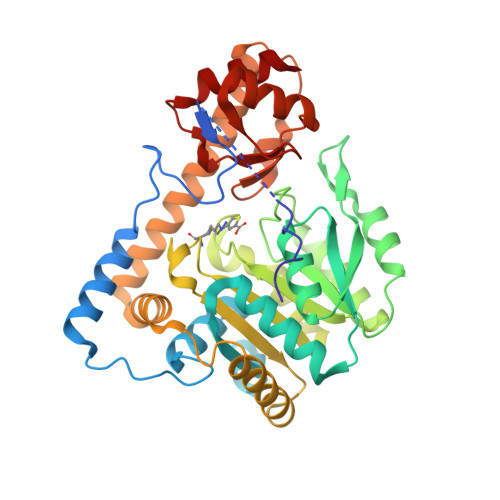
    As a mechanism-based inactivator of PLP-enzymes, (S)-4-amino-4,5-dihydro-2-thiophenecarboxylic acid (SADTA) was cocrystallized with Escherichia coli aspartate aminotransferase (l-AspAT) at a series of pH values ranging from 6 to 8. Five structural models with high resolution (1.4-1.85 A) were obtained for l-AspAT-SADTA complexes at pH 6.0, 6.5, 7.0, 7.5, and 8.0. Electron densities of the models showed that two different adducts had formed in the active sites. One adduct was formed from SADTA covalently linked to pyridoxal 5'-phosphate (PLP) while the other adduct was formed with the inhibitor covalently linked to Lysine246,1 the active site lysine. Moreover, there is a strong indication based on the electron densities that the occurrence of the two adducts is pH dependent. We conclude that SADTA inactivates l-AspAT via two different mechanisms based on the binding direction of the inactivator. Additionally, the structural models also show pH dependence of the protein structure itself, which provided detailed mechanistic implications for l-AspAT.


  • Organizational Affiliation
    • Department of Biochemistry and Chemistry and Rosenstiel Basic Medical Sciences Research Center MS029, Brandeis University, Waltham, Massachusetts 02454-9110, USA.

Macromolecule Content 

  • Total Structure Weight: 45.48 kDa 
  • Atom Count: 3,808 
  • Modeled Residue Count: 379 
  • Deposited Residue Count: 396 
  • Unique protein chains: 1

Macromolecules

Find similar proteins by:|  3D Structure
Entity ID: 1
MoleculeChains  Sequence LengthOrganismDetailsImage
Aspartate aminotransferase396Escherichia coliMutation(s): 1 
Gene Names: aspC
EC: 2.6.1.1
UniProt
Find proteins for P00509 (Escherichia coli (strain K12))
Explore P00509 
Go to UniProtKB:  P00509
Entity Groups
Sequence Clusters30% Identity50% Identity70% Identity90% Identity95% Identity100% Identity
UniProt GroupP00509
Sequence Annotations
Expand
Reference Sequence

Small Molecules

Ligands 4 Unique
IDChains Name / Formula / InChI Key2D Diagram3D Interactions
PSZ

Query on PSZ


Download:Ideal Coordinates CCD File
C [auth A]4-[({3-HYDROXY-2-METHYL-5-[(PHOSPHONOOXY)METHYL]PYRIDIN-4-YL}METHYL)AMINO]THIOPHENE-2-CARBOXYLIC ACID
C13 H15 N2 O7 P S
SUKBSASAEROGCJ-UHFFFAOYSA-N
PMP

Query on PMP


Download:Ideal Coordinates CCD File
D [auth A]4'-DEOXY-4'-AMINOPYRIDOXAL-5'-PHOSPHATE
C8 H13 N2 O5 P
ZMJGSOSNSPKHNH-UHFFFAOYSA-N
SO4

Query on SO4


Download:Ideal Coordinates CCD File
B [auth A]SULFATE ION
O4 S
QAOWNCQODCNURD-UHFFFAOYSA-L
GOL

Query on GOL


Download:Ideal Coordinates CCD File
E [auth A]
F [auth A]
G [auth A]
H [auth A]
I [auth A]
E [auth A],
F [auth A],
G [auth A],
H [auth A],
I [auth A],
J [auth A],
K [auth A],
L [auth A],
M [auth A],
N [auth A],
O [auth A]
GLYCEROL
C3 H8 O3
PEDCQBHIVMGVHV-UHFFFAOYSA-N
Modified Residues  1 Unique
IDChains TypeFormula2D DiagramParent
KST
Query on KST
A
L-PEPTIDE LINKINGC11 H16 N2 O4 SLYS

Experimental Data & Validation

Experimental Data

  • Method: X-RAY DIFFRACTION
  • Resolution: 1.40 Å
  • R-Value Free:  0.188 (Depositor), 0.187 (DCC) 
  • R-Value Work:  0.154 (Depositor), 0.152 (DCC) 
  • R-Value Observed: 0.156 (Depositor) 
Space Group: C 2 2 21
Unit Cell:
Length ( Å )Angle ( ˚ )
a = 153.623α = 90
b = 85.135β = 90
c = 78.849γ = 90
Software Package:
Software NamePurpose
REFMACrefinement
ADSCdata collection
HKL-2000data reduction
HKL-2000data scaling
PHASERphasing

Structure Validation

View Full Validation Report


Ligand Structure Quality Assessment 


Entry History 

Deposition Data

Revision History  (Full details and data files)

  • Version 1.0: 2007-09-04
    Type: Initial release
  • Version 1.1: 2011-07-13
    Changes: Derived calculations, Version format compliance
  • Version 1.2: 2023-08-30
    Changes: Data collection, Database references, Derived calculations, Refinement description