2Q71 | pdb_00002q71

Uroporphyrinogen Decarboxylase G168R single mutant enzyme in complex with coproporphyrinogen-III


Experimental Data Snapshot

  • Method: X-RAY DIFFRACTION
  • Resolution: 1.90 Å
  • R-Value Free: 
    0.199 (Depositor), 0.202 (DCC) 
  • R-Value Work: 
    0.162 (Depositor), 0.164 (DCC) 
  • R-Value Observed: 
    0.164 (Depositor) 

Starting Model: experimental
View more details

wwPDB Validation 3D Report Full Report

Validation slider image for 2Q71

Ligand Structure Quality Assessment 


This is version 1.6 of the entry. See complete history

Literature

Two Novel Uropophyrinogen Decarboxylase (URO-D) Mutations Causing Hepatoerythropoietic Porphyria (HEP)

Phillips, J.D.Whitby, F.G.Stadtmueller, B.M.Edwards, C.Q.Hill, C.P.Kushner, J.P.

(2007) Transl Res 149: 85-91

  • DOI: https://doi.org/10.1016/j.trsl.2006.08.006
  • Primary Citation Related Structures: 
    2Q6Z, 2Q71

  • PubMed Abstract: 

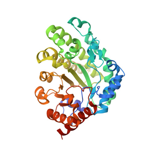
    Hepatoerythropoietic porphyria (HEP) is a rare form of porphyria in humans. The disorder is caused by homozygosity or compound heterozygosity for mutations of the uroporphyrinogen decarboxylase (URO-D) gene. Subnormal URO-D activity results in accumulation of uroporphyrin in the liver, which ultimately mediates the photosensitivity that clinically characterizes HEP. Two previously undescribed URO-D mutations found in a 2-year-old Caucasian boy with HEP, a maternal nonsense mutation (Gln71Stop), and a paternal missense mutation (Gly168Arg) are reported here. Recombinant Gly168Arg URO-D retained 65% of wild-type URO-D activity and studies in Epstein-Barr Virus (EBV)-transformed lymphoblasts indicated that protein levels are reduced, suggesting that the mutant protein might be subjected to accelerated turnover. The crystal structure of Gly168Arg was determined both as the apo-enzyme and with the reaction product bound. These studies revealed little distortion of the active site, but a loop containing residues 167-172 was displaced, possibly indicating small changes in the catalytic geometry or in substrate binding or increased accessibility to a cellular proteolytic pathway. A second pregnancy occurred in this family, and in utero genotyping revealed a fetus heterozygous for the maternal nonsense mutation (URO-D genotype WT/Gln71Stop). A healthy infant was born with no clinical evidence of porphyria.


  • Organizational Affiliation
    • Department of Medicine, University of Utah School of Medicine, Salt Lake City, UT 84132, USA. john.phillips@hsc.utah.edu

Macromolecule Content 

  • Total Structure Weight: 40.52 kDa 
  • Atom Count: 3,428 
  • Modeled Residue Count: 356 
  • Deposited Residue Count: 356 
  • Unique protein chains: 1

Macromolecules

Find similar proteins by:|  3D Structure
Entity ID: 1
MoleculeChains  Sequence LengthOrganismDetailsImage
Uroporphyrinogen decarboxylase356Homo sapiensMutation(s): 1 
Gene Names: UROD
EC: 4.1.1.37
UniProt & NIH Common Fund Data Resources
Find proteins for P06132 (Homo sapiens)
Explore P06132 
Go to UniProtKB:  P06132
PHAROS:  P06132
GTEx:  ENSG00000126088 
Entity Groups
Sequence Clusters30% Identity50% Identity70% Identity90% Identity95% Identity100% Identity
UniProt GroupP06132
Sequence Annotations
Expand
Reference Sequence

Small Molecules

Ligands 1 Unique
IDChains Name / Formula / InChI Key2D Diagram3D Interactions
CP3

Query on CP3


Download:Ideal Coordinates CCD File
B [auth A]COPROPORPHYRINOGEN III
C36 H44 N4 O8
NIUVHXTXUXOFEB-UHFFFAOYSA-N

Experimental Data & Validation

Experimental Data

  • Method: X-RAY DIFFRACTION
  • Resolution: 1.90 Å
  • R-Value Free:  0.199 (Depositor), 0.202 (DCC) 
  • R-Value Work:  0.162 (Depositor), 0.164 (DCC) 
  • R-Value Observed: 0.164 (Depositor) 
Space Group: P 31 2 1
Unit Cell:
Length ( Å )Angle ( ˚ )
a = 103.037α = 90
b = 103.037β = 90
c = 71.865γ = 120
Software Package:
Software NamePurpose
DENZOdata reduction
SCALEPACKdata scaling
REFMACrefinement
PDB_EXTRACTdata extraction
REFMACphasing

Structure Validation

View Full Validation Report


Ligand Structure Quality Assessment 


Entry History 

Deposition Data

Revision History  (Full details and data files)

  • Version 1.0: 2007-06-19
    Type: Initial release
  • Version 1.1: 2008-05-01
    Changes: Version format compliance
  • Version 1.2: 2011-07-13
    Changes: Version format compliance
  • Version 1.3: 2014-03-12
    Changes: Non-polymer description
  • Version 1.4: 2017-10-18
    Changes: Refinement description
  • Version 1.5: 2021-10-20
    Changes: Database references, Derived calculations
  • Version 1.6: 2023-08-30
    Changes: Data collection, Refinement description