2Q66 | pdb_00002q66

Structure of Yeast Poly(A) Polymerase with ATP and oligo(A)


Experimental Data Snapshot

  • Method: X-RAY DIFFRACTION
  • Resolution: 1.80 Å
  • R-Value Free: 
    0.225 (Depositor), 0.223 (DCC) 
  • R-Value Work: 
    0.189 (Depositor), 0.187 (DCC) 
  • R-Value Observed: 
    0.191 (Depositor) 

Starting Model: experimental
View more details

wwPDB Validation 3D Report Full Report

Validation slider image for 2Q66

Ligand Structure Quality Assessment 


This is version 1.4 of the entry. See complete history

Literature

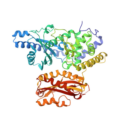
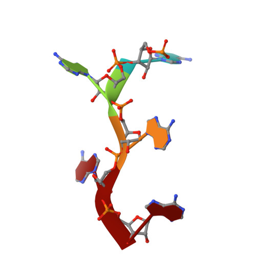
Mechanism of poly(A) polymerase: structure of the enzyme-MgATP-RNA ternary complex and kinetic analysis.

Balbo, P.B.Bohm, A.

(2007) Structure 15: 1117-1131

  • DOI: https://doi.org/10.1016/j.str.2007.07.010
  • Primary Citation Related Structures: 
    2Q66

  • PubMed Abstract: 

    We report the 1.8 A structure of yeast poly(A) polymerase (PAP) trapped in complex with ATP and a five residue poly(A) by mutation of the catalytically required aspartic acid 154 to alanine. The enzyme has undergone significant domain movement and reveals a closed conformation with extensive interactions between the substrates and all three polymerase domains. Both substrates and 31 buried water molecules are enclosed within a central cavity that is open at both ends. Four PAP mutants were subjected to detailed kinetic analysis, and studies of the adenylyltransfer (forward), pyrophosphorolysis (reverse), and nucleotidyltransfer reaction utilizing CTP for the mutants are presented. The results support a model in which binding of both poly(A) and the correct nucleotide, MgATP, induces a conformational change, resulting in formation of a stable, closed enzyme state. Thermodynamic considerations of the data are discussed as they pertain to domain closure, substrate specificity, and catalytic strategies utilized by PAP.


  • Organizational Affiliation
    • Department of Biochemistry, Tufts School of Medicine, 136 Harrison Avenue, Boston, MA 02111, USA.

Macromolecule Content 

  • Total Structure Weight: 63.19 kDa 
  • Atom Count: 5,049 
  • Modeled Residue Count: 524 
  • Deposited Residue Count: 530 
  • Unique protein chains: 1
  • Unique nucleic acid chains: 1

Macromolecules


Find similar proteins by:|  3D Structure
Entity ID: 2
MoleculeChains  Sequence LengthOrganismDetailsImage
Poly(A) polymeraseB [auth A]525Saccharomyces cerevisiaeMutation(s): 1 
Gene Names: PAP1
EC: 2.7.7.19
UniProt
Find proteins for P29468 (Saccharomyces cerevisiae (strain ATCC 204508 / S288c))
Explore P29468 
Go to UniProtKB:  P29468
Entity Groups
Sequence Clusters30% Identity50% Identity70% Identity90% Identity95% Identity100% Identity
UniProt GroupP29468
Sequence Annotations
Expand
Reference Sequence
Find similar nucleic acids by:  Sequence
Entity ID: 1
MoleculeChains LengthOrganismImage
5'-R(P*AP*AP*AP*AP*A)-3'A [auth X]5N/A
Sequence Annotations
Expand
Reference Sequence

Small Molecules

Ligands 3 Unique
IDChains Name / Formula / InChI Key2D Diagram3D Interactions
ATP

Query on ATP


Download:Ideal Coordinates CCD File
F [auth A]ADENOSINE-5'-TRIPHOSPHATE
C10 H16 N5 O13 P3
ZKHQWZAMYRWXGA-KQYNXXCUSA-N
EDO

Query on EDO


Download:Ideal Coordinates CCD File
C [auth X]
D [auth X]
G [auth A]
H [auth A]
I [auth A]
C [auth X],
D [auth X],
G [auth A],
H [auth A],
I [auth A],
J [auth A],
K [auth A],
L [auth A],
M [auth A],
N [auth A],
O [auth A],
P [auth A],
Q [auth A],
R [auth A],
S [auth A],
T [auth A],
U [auth A]
1,2-ETHANEDIOL
C2 H6 O2
LYCAIKOWRPUZTN-UHFFFAOYSA-N
MG

Query on MG


Download:Ideal Coordinates CCD File
E [auth A]MAGNESIUM ION
Mg
JLVVSXFLKOJNIY-UHFFFAOYSA-N

Experimental Data & Validation

Experimental Data

  • Method: X-RAY DIFFRACTION
  • Resolution: 1.80 Å
  • R-Value Free:  0.225 (Depositor), 0.223 (DCC) 
  • R-Value Work:  0.189 (Depositor), 0.187 (DCC) 
  • R-Value Observed: 0.191 (Depositor) 
Space Group: P 21 21 21
Unit Cell:
Length ( Å )Angle ( ˚ )
a = 67.633α = 90
b = 85.907β = 90
c = 107.469γ = 90
Software Package:
Software NamePurpose
DENZOdata reduction
SCALEPACKdata scaling
REFMACrefinement
PDB_EXTRACTdata extraction
ADSCdata collection
MOLREPphasing

Structure Validation

View Full Validation Report


Ligand Structure Quality Assessment 


Entry History 

Deposition Data

Revision History  (Full details and data files)

  • Version 1.0: 2007-08-28
    Type: Initial release
  • Version 1.1: 2011-07-13
    Changes: Version format compliance
  • Version 1.2: 2017-10-18
    Changes: Refinement description
  • Version 1.3: 2021-10-20
    Changes: Database references, Derived calculations
  • Version 1.4: 2023-08-30
    Changes: Data collection, Refinement description