2Q5G | pdb_00002q5g

Ligand binding domain of PPAR delta receptor in complex with a partial agonist


Experimental Data Snapshot

  • Method: X-RAY DIFFRACTION
  • Resolution: 2.70 Å
  • R-Value Free: 
    0.305 (Depositor), 0.302 (DCC) 
  • R-Value Work: 
    0.225 (Depositor), 0.231 (DCC) 
  • R-Value Observed: 
    0.229 (Depositor) 

Starting Model: experimental
View more details

wwPDB Validation 3D Report Full Report

Validation slider image for 2Q5G

Ligand Structure Quality Assessment 


This is version 1.3 of the entry. See complete history

Literature

Design of a partial PPARdelta agonist.

Pettersson, I.Ebdrup, S.Havranek, M.Pihera, P.Korinek, M.Mogensen, J.P.Jeppesen, C.B.Johansson, E.Sauerberg, P.

(2007) Bioorg Med Chem Lett 17: 4625-4629

  • DOI: https://doi.org/10.1016/j.bmcl.2007.05.079
  • Primary Citation Related Structures: 
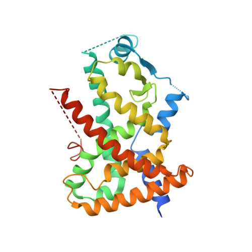
    2Q5G

  • PubMed Abstract: 

    Structure based ligand design was used in order to design a partial agonist for the PPARdelta receptor. The maximum activation in the transactivation assay was reduced from 87% to 39%. The crystal structure of the ligand binding domain of the PPARdelta receptor in complex with compound 2 was determined in order to understand the structural changes which gave rise to the decrease in maximum activation.


  • Organizational Affiliation
    • Novo Nordisk A/S, Novo Nordisk Park, 2760 Måløv, Denmark. inpe@novonordisk.com

Macromolecule Content 

  • Total Structure Weight: 66.17 kDa 
  • Atom Count: 3,876 
  • Modeled Residue Count: 479 
  • Deposited Residue Count: 566 
  • Unique protein chains: 1

Macromolecules

Find similar proteins by:|  3D Structure
Entity ID: 1
MoleculeChains  Sequence LengthOrganismDetailsImage
Peroxisome proliferator-activated receptor delta
A, B
283Homo sapiensMutation(s): 0 
Gene Names: PPARDNR1C2PPARB
UniProt & NIH Common Fund Data Resources
Find proteins for Q03181 (Homo sapiens)
Explore Q03181 
Go to UniProtKB:  Q03181
PHAROS:  Q03181
GTEx:  ENSG00000112033 
Entity Groups
Sequence Clusters30% Identity50% Identity70% Identity90% Identity95% Identity100% Identity
UniProt GroupQ03181
Sequence Annotations
Expand
Reference Sequence

Small Molecules

Ligands 1 Unique
IDChains Name / Formula / InChI Key2D Diagram3D Interactions
1FA

Query on 1FA


Download:Ideal Coordinates CCD File
C [auth A],
D [auth B]
[(7-{[2-(3-MORPHOLIN-4-YLPROP-1-YN-1-YL)-6-{[4-(TRIFLUOROMETHYL)PHENYL]ETHYNYL}PYRIDIN-4-YL]THIO}-2,3-DIHYDRO-1H-INDEN- 4-YL)OXY]ACETIC ACID
C32 H27 F3 N2 O4 S
AKQAEZPFLKFQCZ-UHFFFAOYSA-N
Binding Affinity Annotations 
IDSourceBinding Affinity
1FA BindingDB:  2Q5G EC50: 130 (nM) from 1 assay(s)

Experimental Data & Validation

Experimental Data

  • Method: X-RAY DIFFRACTION
  • Resolution: 2.70 Å
  • R-Value Free:  0.305 (Depositor), 0.302 (DCC) 
  • R-Value Work:  0.225 (Depositor), 0.231 (DCC) 
  • R-Value Observed: 0.229 (Depositor) 
Space Group: C 1 2 1
Unit Cell:
Length ( Å )Angle ( ˚ )
a = 112.577α = 90
b = 65.603β = 124.05
c = 101.149γ = 90
Software Package:
Software NamePurpose
d*TREKdata scaling
REFMACrefinement
PDB_EXTRACTdata extraction
d*TREKdata reduction
PHASESphasing

Structure Validation

View Full Validation Report


Ligand Structure Quality Assessment 


Entry History 

Deposition Data

Revision History  (Full details and data files)

  • Version 1.0: 2008-06-03
    Type: Initial release
  • Version 1.1: 2011-07-13
    Changes: Derived calculations, Version format compliance
  • Version 1.2: 2018-01-24
    Changes: Structure summary
  • Version 1.3: 2023-08-30
    Changes: Data collection, Database references, Derived calculations, Refinement description, Structure summary