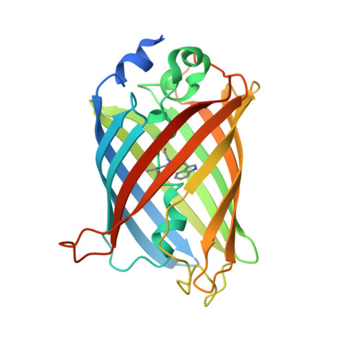
X-ray structure of Cerulean GFP: a tryptophan-based chromophore useful for fluorescence lifetime imaging.
Malo, G.D., Pouwels, L.J., Wang, M., Weichsel, A., Montfort, W.R., Rizzo, M.A., Piston, D.W., Wachter, R.M.(2007) Biochemistry 46: 9865-9873
- PubMed: 17685554 Search on PubMed
- DOI: https://doi.org/10.1021/bi602664c
- Primary Citation Related Structures:
2Q57 - PubMed Abstract:
The crystal structure of the cyan-fluorescent Cerulean green fluorescent protein (GFP), a variant of enhanced cyan fluorescent protein (ECFP), has been determined to 2.0 A. Cerulean bears an internal fluorophore composed of an indole moiety derived from Y66W, conjugated to the GFP-like imidazolinone ring via a methylene bridge. Cerulean undergoes highly efficient fluorescence resonance energy transfer (FRET) to yellow acceptor molecules and exhibits significantly reduced excited-state heterogeneity. This feature was rationally engineered in ECFP by substituting His148 with an aspartic acid [Rizzo et al. (2004) Nat. Biotechnol. 22, 445], rendering Cerulean useful for fluorescence lifetime imaging microscopy (FLIM). The X-ray structure is consistent with a single conformation of the chromophore and surrounding residues and may therefore provide a structural rationale for the previously described monoexponential fluorescence decay. Unexpectedly, the carboxyl group of H148D is found in a buried position, directly contacting the indole nitrogen of the chromophore via a bifurcated hydrogen bond. Compared to the similarly constructed ECFP chromophore, the indole group of Cerulean is rotated around the methylene bridge to adopt a cis-coplanar conformation with respect to the imidazolinone ring, resulting in a close edge-to-edge contact of the two ring systems. The double-humped absorbance spectrum persists in single-crystal absorbance measurements, casting doubt on the idea that ground state conformational heterogeneity forms the basis of the two overlapping transitions. At low pH, a blue shift in absorbance of 10-15 nm suggests a pH-induced structural transition that proceeds with a time constant of 47 (+/-2) min and is reversible. Possible interpretations in terms of chromophore isomerization are presented.
- Department of Chemistry and Biochemistry, Arizona State University, Tempe, Arizona 85287-1604, USA.
Organizational Affiliation: