2Q2L | pdb_00002q2l

Crystal Structure of Superoxide Dismutase from P. atrosanguina


Experimental Data Snapshot

  • Method: X-RAY DIFFRACTION
  • Resolution: 2.37 Å
  • R-Value Free: 
    0.234 (Depositor), 0.221 (DCC) 
  • R-Value Work: 
    0.167 (Depositor) 

wwPDB Validation 3D Report Full Report

Validation slider image for 2Q2L

This is version 1.2 of the entry. See complete history

Literature

SAD phasing of a structure based on cocrystallized iodides using an in-house Cu Kalpha X-ray source: effects of data redundancy and completeness on structure solution

Yogavel, M.Gill, J.Mishra, P.C.Sharma, A.

(2007) Acta Crystallogr D Biol Crystallogr 63: 931-934

  • DOI: https://doi.org/10.1107/S0907444907029174
  • Primary Citation Related Structures: 
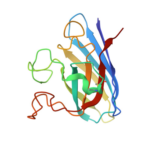
    2Q2L

  • PubMed Abstract: 

    Superoxide dismutase (SOD) from Potentilla atrosanguinea (Wall. ex. Lehm.) was crystallized using 20% PEG 3350 and 0.2 M ammonium iodide and diffraction data were collected to 2.36 A resolution using an in-house Cu Kalpha X-ray source. Analyses show that data with a redundancy of 3.2 were sufficient to determine the structure by the SAD technique using the iodine anomalous signal. This redundancy is lower than that in previous cases in which protein structures were determined using iodines for phasing and in-house copper X-ray sources. Cocrystallization of proteins with halide salts such as ammonium iodide in combination with copper-anode X-ray radiation can therefore serve as a powerful and easy avenue for structure solution.


  • Organizational Affiliation
    • Structural and Computational Biology Group, International Centre for Genetic Engineering and Biotechnology, New Delhi, India.

Macromolecule Content 

  • Total Structure Weight: 31.83 kDa 
  • Atom Count: 2,399 
  • Modeled Residue Count: 304 
  • Deposited Residue Count: 304 
  • Unique protein chains: 1

Macromolecules

Find similar proteins by:|  3D Structure
Entity ID: 1
MoleculeChains  Sequence LengthOrganismDetailsImage
Superoxide Dismutase
A, B
152Potentilla atrosanguineaMutation(s): 0 
EC: 1.15.1.1
UniProt
Find proteins for B2CP37 (Potentilla atrosanguinea)
Explore B2CP37 
Go to UniProtKB:  B2CP37
Entity Groups
Sequence Clusters30% Identity50% Identity70% Identity90% Identity95% Identity100% Identity
UniProt GroupB2CP37
Sequence Annotations
Expand
Reference Sequence

Small Molecules

Ligands 2 Unique
IDChains Name / Formula / InChI Key2D Diagram3D Interactions
IOD

Query on IOD


Download:Ideal Coordinates CCD File
D [auth A]
E [auth A]
F [auth A]
G [auth A]
H [auth A]
D [auth A],
E [auth A],
F [auth A],
G [auth A],
H [auth A],
I [auth A],
K [auth B],
L [auth B],
M [auth B],
N [auth B]
IODIDE ION
I
XMBWDFGMSWQBCA-UHFFFAOYSA-M
ZN

Query on ZN


Download:Ideal Coordinates CCD File
C [auth A],
J [auth B]
ZINC ION
Zn
PTFCDOFLOPIGGS-UHFFFAOYSA-N

Experimental Data & Validation

Experimental Data

  • Method: X-RAY DIFFRACTION
  • Resolution: 2.37 Å
  • R-Value Free:  0.234 (Depositor), 0.221 (DCC) 
  • R-Value Work:  0.167 (Depositor) 
Space Group: C 1 2 1
Unit Cell:
Length ( Å )Angle ( ˚ )
a = 89.14α = 90
b = 64.12β = 124.33
c = 63.62γ = 90
Software Package:
Software NamePurpose
MAR345dtbdata collection
SOLVEphasing
REFMACrefinement
HKL-2000data reduction
HKL-2000data scaling

Structure Validation

View Full Validation Report


Entry History 

Deposition Data

Revision History  (Full details and data files)

  • Version 1.0: 2008-03-25
    Type: Initial release
  • Version 1.1: 2011-07-13
    Changes: Version format compliance
  • Version 1.2: 2024-10-30
    Changes: Data collection, Database references, Derived calculations, Structure summary