


Crystal Structure of Phospholipase A2 complex with propanol at 1.5 A resolution
Kumar, S., Hariprasad, G., Singh, N., Sharma, S., Kaur, P., Perbandt, M., Betzel, C., Singh, T.P.To be published.
Experimental Data Snapshot
Starting Model: experimental
View more details
wwPDB Validation 3D Report Full Report

Entity ID: 1 | |||||
|---|---|---|---|---|---|
| Molecule | Chains | Sequence Length | Organism | Details | Image |
| Phospholipase A2 VRV-PL-VIIIa | 121 | Daboia russelii pulchella | Mutation(s): 0 EC: 3.1.1.4 | 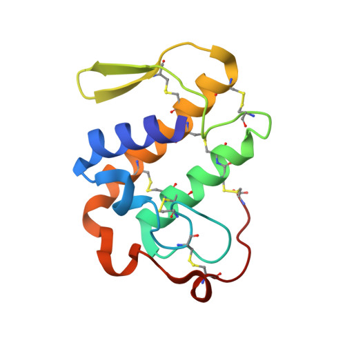 | |
UniProt | |||||
Entity Groups | |||||
| Sequence Clusters | 30% Identity50% Identity70% Identity90% Identity95% Identity100% Identity | ||||
| UniProt Group | P59071 | ||||
Sequence AnnotationsExpand | |||||
Reference Sequence | |||||
| Ligands 2 Unique | |||||
|---|---|---|---|---|---|
| ID | Chains | Name / Formula / InChI Key | 2D Diagram | 3D Interactions | |
| SO4 Download:Ideal Coordinates CCD File | B [auth A], C [auth A], D [auth A], E [auth A] | SULFATE ION O4 S QAOWNCQODCNURD-UHFFFAOYSA-L |  | ||
| POL Download:Ideal Coordinates CCD File | F [auth A] | N-PROPANOL C3 H8 O BDERNNFJNOPAEC-UHFFFAOYSA-N |  | ||
| Length ( Å ) | Angle ( ˚ ) |
|---|---|
| a = 52.15 | α = 90 |
| b = 52.15 | β = 90 |
| c = 47.611 | γ = 90 |
| Software Name | Purpose |
|---|---|
| REFMAC | refinement |
| HKL-2000 | data collection |
| HKL-2000 | data reduction |
| SCALEPACK | data scaling |
| AMoRE | phasing |


