2Q0A | pdb_00002q0a

Structure and rearrangements in the carboxy-terminal region of SpIH channels


Experimental Data Snapshot

  • Method: X-RAY DIFFRACTION
  • Resolution: 2.25 Å
  • R-Value Free: 
    0.261 (Depositor), 0.259 (DCC) 
  • R-Value Work: 
    0.209 (Depositor), 0.206 (DCC) 

Starting Model: experimental
View more details

wwPDB Validation 3D Report Full Report

Validation slider image for 2Q0A

Ligand Structure Quality Assessment 


This is version 1.5 of the entry. See complete history

Literature

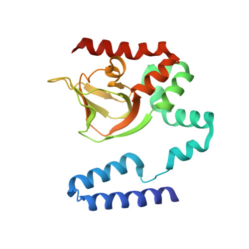
Structure and rearrangements in the carboxy-terminal region of SpIH channels.

Flynn, G.E.Black, K.D.Islas, L.D.Sankaran, B.Zagotta, W.N.

(2007) Structure 15: 671-682

  • DOI: https://doi.org/10.1016/j.str.2007.04.008
  • Primary Citation Related Structures: 
    2PTM, 2Q0A

  • PubMed Abstract: 

    Hyperpolarization-activated cyclic nucleotide-modulated (HCN) ion channels regulate the spontaneous firing activity and electrical excitability of many cardiac and neuronal cells. The modulation of HCN channel opening by the direct binding of cAMP underlies many physiological processes such as the autonomic regulation of the heart rate. Here we use a combination of X-ray crystallography and electrophysiology to study the allosteric mechanism for cAMP modulation of HCN channels. SpIH is an invertebrate HCN channel that is activated fully by cAMP, but only partially by cGMP. We exploited the partial agonist action of cGMP on SpIH to reveal the molecular mechanism for cGMP specificity of many cyclic nucleotide-regulated enzymes. Our results also elaborate a mechanism for the allosteric conformational change in the cyclic nucleotide-binding domain and a mechanism for partial agonist action. These mechanisms will likely extend to other cyclic nucleotide-regulated channels and enzymes as well.


  • Organizational Affiliation
    • Department of Physiology and Biophysics, Howard Hughes Medical Institute, University of Washington, Seattle, WA 98195, USA.

Macromolecule Content 

  • Total Structure Weight: 47.55 kDa 
  • Atom Count: 3,467 
  • Modeled Residue Count: 388 
  • Deposited Residue Count: 400 
  • Unique protein chains: 1

Macromolecules

Find similar proteins by:|  3D Structure
Entity ID: 1
MoleculeChains  Sequence LengthOrganismDetailsImage
Potassium/sodium hyperpolarization-activated cyclic nucleotide-gated channel 2
A, B
200Mus musculusMutation(s): 1 
Gene Names: Hcn2Bcng2Hac1
UniProt
Find proteins for O88703 (Mus musculus)
Explore O88703 
Go to UniProtKB:  O88703
Entity Groups
Sequence Clusters30% Identity50% Identity70% Identity90% Identity95% Identity100% Identity
UniProt GroupO88703
Sequence Annotations
Expand
Reference Sequence

Experimental Data & Validation

Experimental Data

  • Method: X-RAY DIFFRACTION
  • Resolution: 2.25 Å
  • R-Value Free:  0.261 (Depositor), 0.259 (DCC) 
  • R-Value Work:  0.209 (Depositor), 0.206 (DCC) 
Space Group: I 4
Unit Cell:
Length ( Å )Angle ( ˚ )
a = 94.734α = 90
b = 94.734β = 90
c = 124.047γ = 90
Software Package:
Software NamePurpose
DENZOdata reduction
SCALEPACKdata scaling
PHENIXrefinement
PDB_EXTRACTdata extraction
HKL-2000data collection
HKL-2000data reduction
CNSphasing

Structure Validation

View Full Validation Report


Ligand Structure Quality Assessment 


Entry History 

Deposition Data

Revision History  (Full details and data files)

  • Version 1.0: 2007-06-19
    Type: Initial release
  • Version 1.1: 2008-05-01
    Changes: Version format compliance
  • Version 1.2: 2011-07-13
    Changes: Derived calculations, Version format compliance
  • Version 1.3: 2017-10-18
    Changes: Advisory, Refinement description
  • Version 1.4: 2021-10-20
    Changes: Advisory, Database references, Derived calculations
  • Version 1.5: 2023-08-30
    Changes: Data collection, Refinement description