2PZ0 | pdb_00002pz0

Crystal structure of Glycerophosphodiester Phosphodiesterase (GDPD) from T. tengcongensis


Experimental Data Snapshot

  • Method: X-RAY DIFFRACTION
  • Resolution: 1.91 Å
  • R-Value Free: 
    0.236 (Depositor), 0.224 (DCC) 
  • R-Value Work: 
    0.229 (Depositor), 0.220 (DCC) 

Starting Model: experimental
View more details

wwPDB Validation 3D Report Full Report

Validation slider image for 2PZ0

This is version 1.3 of the entry. See complete history

Literature

Crystal structure of glycerophosphodiester phosphodiesterase (GDPD) from Thermoanaerobacter tengcongensis, a metal ion-dependent enzyme: insight into the catalytic mechanism.

Shi, L.Liu, J.F.An, X.M.Liang, D.C.

(2008) Proteins 72: 280-288

  • DOI: https://doi.org/10.1002/prot.21921
  • Primary Citation Related Structures: 
    2PZ0

  • PubMed Abstract: 

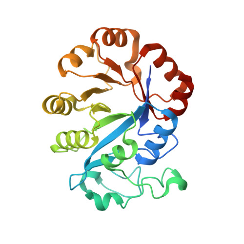
    Glycerophosphodiester phosphodiesterase (GDPD; EC 3.1.4.46) catalyzes the hydrolysis of a glycerophosphodiester to an alcohol and glycerol 3-phosphate in glycerol metabolism. It has an important role in the synthesis of a variety of products that participate in many biochemical pathways. We report the crystal structure of the Thermoanaerobacter tengcongensis GDPD (ttGDPD) at 1.91 A resolution, with a calcium ion and glycerol as a substrate mimic coordinated at this calcium ion (PDB entry 2pz0). The ttGDPD dimer with an intermolecular disulfide bridge and two hydrogen bonds is considered as the potential functional unit. We used site-directed mutagenesis to characterize ttGDPD as a metal ion-dependent enzyme, identified a cluster of residues involved in substrate binding and the catalytic reaction, and we propose a possible general acid-base catalytic mechanism for ttGDPD. Superposing the active site with the homologous structure GDPD from Agrobacterium tumefaciens (PDB entry 1zcc), which binds a sulfate ion in the active site, the sulfate ion can represent the phosphate moiety of the substrate, simulating the binding mode of the true substrate of GDPD.


  • Organizational Affiliation
    • National Laboratory of Biomacromolecules, Institute of Biophysics, Chinese Academy of Sciences, Beijing 100101, People's Republic of China.

Macromolecule Content 

  • Total Structure Weight: 57.83 kDa 
  • Atom Count: 4,209 
  • Modeled Residue Count: 482 
  • Deposited Residue Count: 504 
  • Unique protein chains: 1

Macromolecules

Find similar proteins by:|  3D Structure
Entity ID: 1
MoleculeChains  Sequence LengthOrganismDetailsImage
Glycerophosphoryl diester phosphodiesterase
A, B
252Caldanaerobacter subterraneus subsp. tengcongensis MB4Mutation(s): 0 
EC: 3.1.4.46
UniProt
Find proteins for Q8RB32 (Caldanaerobacter subterraneus subsp. tengcongensis (strain DSM 15242 / JCM 11007 / NBRC 100824 / MB4))
Explore Q8RB32 
Go to UniProtKB:  Q8RB32
Entity Groups
Sequence Clusters30% Identity50% Identity70% Identity90% Identity95% Identity100% Identity
UniProt GroupQ8RB32
Sequence Annotations
Expand
Reference Sequence

Experimental Data & Validation

Experimental Data

  • Method: X-RAY DIFFRACTION
  • Resolution: 1.91 Å
  • R-Value Free:  0.236 (Depositor), 0.224 (DCC) 
  • R-Value Work:  0.229 (Depositor), 0.220 (DCC) 
Space Group: P 1 21 1
Unit Cell:
Length ( Å )Angle ( ˚ )
a = 49.401α = 90
b = 53.608β = 97.91
c = 110.885γ = 90
Software Package:
Software NamePurpose
CrystalCleardata collection
PHASERphasing
CNSrefinement
DENZOdata reduction
SCALEPACKdata scaling

Structure Validation

View Full Validation Report


Entry History 

Deposition Data

Revision History  (Full details and data files)

  • Version 1.0: 2008-04-01
    Type: Initial release
  • Version 1.1: 2011-07-13
    Changes: Source and taxonomy, Version format compliance
  • Version 1.2: 2023-08-30
    Changes: Data collection, Database references, Derived calculations, Refinement description
  • Version 1.3: 2024-11-20
    Changes: Structure summary