2PY9

Protein-RNA Interaction involving KH1 domain from Human Poly(C)-Binding Protein-2


Experimental Data Snapshot

  • Method: X-RAY DIFFRACTION
  • Resolution: 2.56 Å
  • R-Value Free: 0.269 
  • R-Value Work: 0.218 
  • R-Value Observed: 0.220 

wwPDB Validation   3D Report Full Report


This is version 1.2 of the entry. See complete history


Literature

X-ray crystallographic and NMR studies of protein-protein and protein-nucleic acid interactions involving the KH domains from human poly(C)-binding protein-2.

Du, Z.Lee, J.K.Fenn, S.Tjhen, R.Stroud, R.M.James, T.L.

(2007) RNA 13: 1043-1051

  • DOI: https://doi.org/10.1261/rna.410107
  • Primary Citation of Related Structures:  
    2PQU, 2PY9

  • PubMed Abstract: 

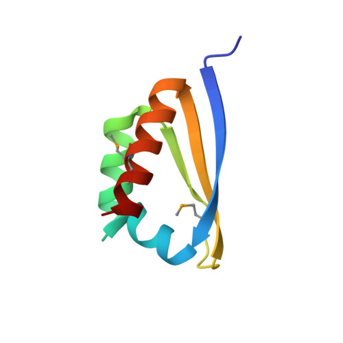
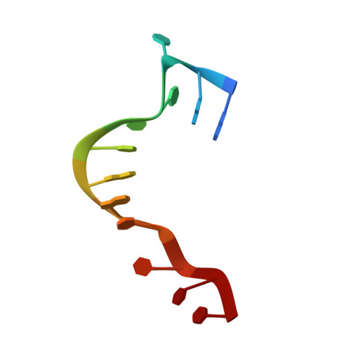
    Poly(C)-binding proteins (PCBPs) are KH (hnRNP K homology) domain-containing proteins that recognize poly(C) DNA and RNA sequences in mammalian cells. Binding poly(C) sequences via the KH domains is critical for PCBP functions. To reveal the mechanisms of KH domain-D/RNA recognition and its functional importance, we have determined the crystal structures of PCBP2 KH1 domain in complex with a 12-nucleotide DNA corresponding to two repeats of the human C-rich strand telomeric DNA and its RNA equivalent. The crystal structures reveal molecular details for not only KH1-DNA/RNA interaction but also protein-protein interaction between two KH1 domains. NMR studies on a protein construct containing two KH domains (KH1 + KH2) of PCBP2 indicate that KH1 interacts with KH2 in a way similar to the KH1-KH1 interaction. The crystal structures and NMR data suggest possible ways by which binding certain nucleic acid targets containing tandem poly(C) motifs may induce structural rearrangement of the KH domains in PCBPs; such structural rearrangement may be crucial for some PCBP functions.


  • Organizational Affiliation

    Department of Pharmaceutical Chemistry, University of California at San Francisco, San Francisco, CA 94158-2517, USA.


Macromolecules

Find similar proteins by:  (by identity cutoff)  |  3D Structure
Entity ID: 2
MoleculeChains Sequence LengthOrganismDetailsImage
Poly(rC)-binding protein 2C [auth A],
D [auth B],
E [auth C],
F [auth D]
73Homo sapiensMutation(s): 3 
Gene Names: PCBP2
UniProt & NIH Common Fund Data Resources
Find proteins for Q15366 (Homo sapiens)
Explore Q15366 
Go to UniProtKB:  Q15366
PHAROS:  Q15366
GTEx:  ENSG00000197111 
Entity Groups  
Sequence Clusters30% Identity50% Identity70% Identity90% Identity95% Identity100% Identity
UniProt GroupQ15366
Sequence Annotations
Expand
  • Reference Sequence

Find similar nucleic acids by:  Sequence   |   3D Structure  

Entity ID: 1
MoleculeChains LengthOrganismImage
12-mer C-rich strand of human telomeric RNAA [auth E],
B [auth F]
12N/A
Sequence Annotations
Expand
  • Reference Sequence
Small Molecules
Modified Residues  1 Unique
IDChains TypeFormula2D DiagramParent
MSE
Query on MSE
C [auth A],
D [auth B],
E [auth C],
F [auth D]
L-PEPTIDE LINKINGC5 H11 N O2 SeMET
Experimental Data & Validation

Experimental Data

  • Method: X-RAY DIFFRACTION
  • Resolution: 2.56 Å
  • R-Value Free: 0.269 
  • R-Value Work: 0.218 
  • R-Value Observed: 0.220 
  • Space Group: P 21 21 2
Unit Cell:
Length ( Å )Angle ( ˚ )
a = 92.006α = 90
b = 58.697β = 90
c = 71.496γ = 90
Software Package:
Software NamePurpose
REFMACrefinement
Blu-Icedata collection
MOSFLMdata reduction
SCALAdata scaling
CNSphasing

Structure Validation

View Full Validation Report


Entry History 

Deposition Data

Revision History  (Full details and data files)

  • Version 1.0: 2007-06-12
    Type: Initial release
  • Version 1.1: 2008-05-01
    Changes: Version format compliance
  • Version 1.2: 2011-07-13
    Changes: Version format compliance