Crystal structure of the EF-hand parvalbumin at atomic resolution (0.91 A) and at low temperature (100 K). Evidence for conformational multistates within the hydrophobic core.
Declercq, J.P., Evrard, C., Lamzin, V., Parello, J.(1999) Protein Sci 8: 2194-2204
- PubMed: 10548066 Search on PubMedSearch on PubMed Central
- DOI: https://doi.org/10.1110/ps.8.10.2194
- Primary Citation Related Structures:
2PVB - PubMed Abstract:
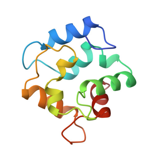
Several crystal structures of parvalbumin (Parv), a typical EF-hand protein, have been reported so far for different species with the best resolution achieving 1.5 A. Using a crystal grown under microgravity conditions, cryotechniques (100 K), and synchrotron radiation, it has now been possible to determine the crystal structure of the fully Ca2+-loaded form of pike (component pI 4.10) Parv.Ca2 at atomic resolution (0.91 A). The availability of such a high quality structure offers the opportunity to contribute to the definition of the validation tools useful for the refinement of protein crystal structures determined to lower resolution. Besides a better definition of most of the elements in the protein three-dimensional structure than in previous studies, the high accuracy thus achieved allows the detection of well-defined alternate conformations, which are observed for 16 residues out of 107 in total. Among them, six occupy an internal position within the hydrophobic core and converge toward two small buried cavities with a total volume of about 60 A3. There is no indication of any water molecule present in these cavities. It is probable that at temperatures of physiological conditions there is a dynamic interconversion between these alternate conformations in an energy-barrier dependent manner. Such motions for which the amplitudes are provided by the present study will be associated with a time-dependent remodeling of the void internal space as part of a slow dynamics regime (millisecond timescales) of the parvalbumin molecule. The relevance of such internal dynamics to function is discussed.
- Université Catholique de Louvain, Unité CPMC, Louvain-la-Neuve, Belgium. declercq@cpmc.ucl.ac.be
Organizational Affiliation: