2PSR | pdb_00002psr

HUMAN PSORIASIN (S100A7) CA2+ AND ZN2+ BOUND FORM (CRYSTAL FORM II)


Experimental Data Snapshot

  • Method: X-RAY DIFFRACTION
  • Resolution: 2.05 Å
  • R-Value Free: 
    0.265 (Depositor), 0.209 (DCC) 
  • R-Value Work: 
    0.187 (DCC) 
  • R-Value Observed: 
    0.215 (Depositor) 

Starting Model: experimental
View more details

wwPDB Validation 3D Report Full Report

Validation slider image for 2PSR

This is version 1.4 of the entry. See complete history

Literature

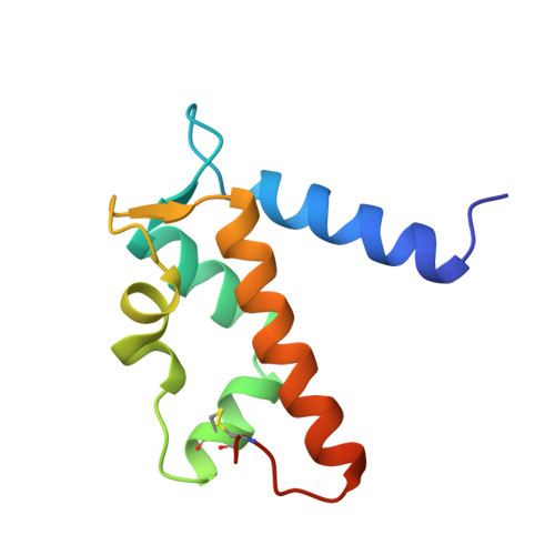
Zinc-binding site of an S100 protein revealed. Two crystal structures of Ca2+-bound human psoriasin (S100A7) in the Zn2+-loaded and Zn2+-free states.

Brodersen, D.E.Nyborg, J.Kjeldgaard, M.

(1999) Biochemistry 38: 1695-1704

  • DOI: https://doi.org/10.1021/bi982483d
  • Primary Citation Related Structures: 
    2PSR, 3PSR

  • PubMed Abstract: 

    The crystal structure of human psoriasin (S100A7) in the native, calcium-bound form has been determined from two crystal forms of the protein crystallized with and without divalent zinc. The overall structures of the dimeric protein closely resemble the previously determined holmium-substituted structure. The structures also reveal a zinc-binding site of the protein, which is formed by three histidines and an aspartate residue. Together, these residues coordinate the zinc ion in a way similar to the pattern seen in certain metalloproteases and in particular the collagenase family of proteins. Sequence comparison suggests that this zinc site is present in a number of the remaining members of the S100 family. The structure of S100A7 crystallized in the absence of zinc further shows that loss of zinc results in a reorganization of the adjacent empty and distorted EF-hand loop, causing it to resemble a calcium-loaded EF-hand.


  • Organizational Affiliation
    • Macromolecular Crystallography, Institute of Molecular and Structural Biology, Aarhus University, Denmark.

Macromolecule Content 

  • Total Structure Weight: 11.45 kDa 
  • Atom Count: 877 
  • Modeled Residue Count: 96 
  • Deposited Residue Count: 100 
  • Unique protein chains: 1

Macromolecules

Find similar proteins by:|  3D Structure
Entity ID: 1
MoleculeChains  Sequence LengthOrganismDetailsImage
PSORIASIN100Homo sapiensMutation(s): 0 
UniProt & NIH Common Fund Data Resources
Find proteins for P31151 (Homo sapiens)
Explore P31151 
Go to UniProtKB:  P31151
PHAROS:  P31151
GTEx:  ENSG00000143556 
Entity Groups
Sequence Clusters30% Identity50% Identity70% Identity90% Identity95% Identity100% Identity
UniProt GroupP31151
Sequence Annotations
Expand
Reference Sequence

Experimental Data & Validation

Experimental Data

  • Method: X-RAY DIFFRACTION
  • Resolution: 2.05 Å
  • R-Value Free:  0.265 (Depositor), 0.209 (DCC) 
  • R-Value Work:  0.187 (DCC) 
  • R-Value Observed: 0.215 (Depositor) 
Space Group: P 43 21 2
Unit Cell:
Length ( Å )Angle ( ˚ )
a = 51.958α = 90
b = 51.958β = 90
c = 116.027γ = 90
Software Package:
Software NamePurpose
DENZOdata reduction
SCALEPACKdata scaling
AMoREphasing
SHELXL-97refinement

Structure Validation

View Full Validation Report


Entry History 

Deposition Data

Revision History  (Full details and data files)

  • Version 1.0: 1999-06-15
    Type: Initial release
  • Version 1.1: 2008-03-25
    Changes: Version format compliance
  • Version 1.2: 2011-07-13
    Changes: Derived calculations, Version format compliance
  • Version 1.3: 2023-08-09
    Changes: Data collection, Database references, Derived calculations, Refinement description
  • Version 1.4: 2024-11-20
    Changes: Data collection, Structure summary