Molecular Architecture of DesI: A Key Enzyme in the Biosynthesis of Desosamine
Burgie, E.S., Holden, H.M.(2007) Biochemistry 46: 8999-9006
- PubMed: 17630700 Search on PubMedSearch on PubMed Central
- DOI: https://doi.org/10.1021/bi700751d
- Primary Citation Related Structures:
2PO3 - PubMed Abstract:
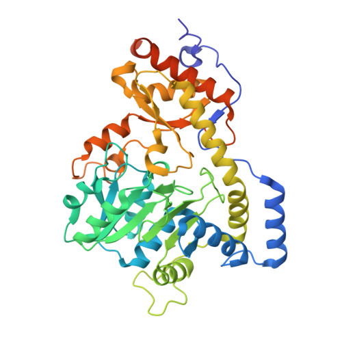
Desosamine is a 3-(dimethylamino)-3,4,6-trideoxyhexose found, for example, in such macrolide antibiotics as erthyromycin, azithromycin, and clarithromycin. The efficacies of these macrolide antibiotics are markedly reduced in the absence of desosamine. In the bacterium Streptomyces venezuelae, six enzymes are required for the production of dTDP-desosamine. The focus of this X-ray crystallographic analysis is the third enzyme in the pathway, a PLP-dependent aminotransferase referred to as DesI. The structure of DesI was solved in complex with its product, dTDP-4-amino-4,6-dideoxyglucose, to a nominal resolution of 2.1 A. Each subunit of the dimeric enzyme contains 12 alpha-helices and 14 beta-strands. Three cis-peptides are observed in each subunit, Phe 330, Pro 332, and Pro 339. The two active sites of the enzyme are located in clefts at the subunit/subunit interface. Electron density corresponding to the bound product clearly demonstrates a covalent bond between the amino group of the product and C-4' of the PLP cofactor. Interestingly, there are no hydrogen-bonding interactions between the protein and the dideoxyglucosyl group of the product (within 3.2 A). The only other sugar-modifying aminotransferase whose structure is known in the presence of product is PseC from Helicobacter pylori. This enzyme, as opposed to DesI, catalyzes amino transfer to the axial position of the sugar. A superposition of the two active sites for these proteins reveals that the major differences in ligand binding occur in the orientations of the deoxyglucosyl and phosphoryl groups. Indeed, the nearly 180 degrees difference in hexose orientation explains the equatorial versus axial amino transfer exhibited by DesI and PseC, respectively.
- Department of Biochemistry, University of Wisconsin, Madison, Wisconsin 53706, USA.
Organizational Affiliation: