2PMI | pdb_00002pmi

Structure of the Pho85-Pho80 CDK-cyclin Complex of the Phosphate-responsive Signal Transduction Pathway with Bound ATP-gamma-S


Experimental Data Snapshot

  • Method: X-RAY DIFFRACTION
  • Resolution: 2.90 Å
  • R-Value Free: 
    0.325 (Depositor), 0.325 (DCC) 
  • R-Value Work: 
    0.289 (Depositor), 0.289 (DCC) 
  • R-Value Observed: 
    0.289 (Depositor) 

Starting Model: experimental
View more details

wwPDB Validation 3D Report Full Report

Validation slider image for 2PMI

Ligand Structure Quality Assessment 


This is version 1.3 of the entry. See complete history

Literature

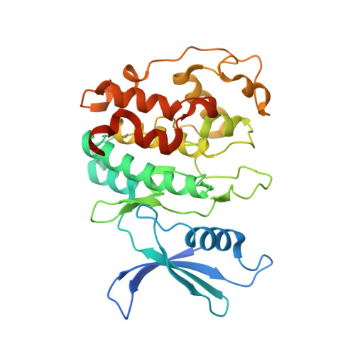
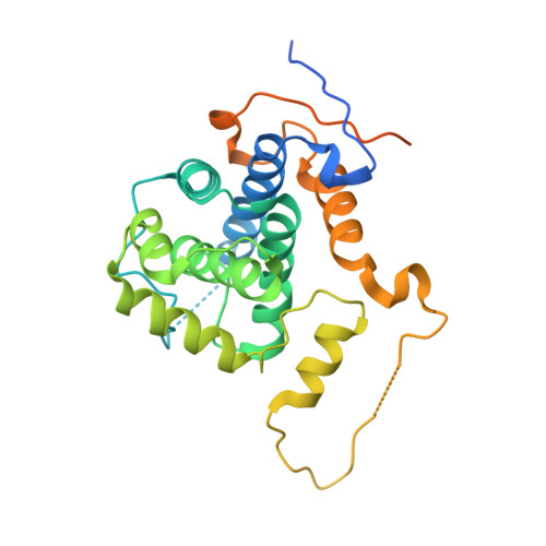
Structure of the Pho85-Pho80 CDK-Cyclin Complex of the Phosphate-Responsive Signal Transduction Pathway

Huang, K.Ferrin-O'Connell, I.Zhang, W.Leonard, G.A.O'Shea, E.K.Quiocho, F.A.

(2007) Mol Cell 28: 614-623

  • DOI: https://doi.org/10.1016/j.molcel.2007.09.013
  • Primary Citation Related Structures: 
    2PK9, 2PMI

  • PubMed Abstract: 

    The ability to sense and respond appropriately to environmental changes is a primary requirement of all living organisms. In response to phosphate limitation, Saccharomyces cerevisiae induces transcription of a set of genes involved in the regulation of phosphate acquisition from the ambient environment. A signal transduction pathway (the PHO pathway) mediates this response, with Pho85-Pho80 playing a vital role. Here we report the X-ray structure of Pho85-Pho80, a prototypic structure of a CDK-cyclin complex functioning in transcriptional regulation in response to environmental changes. The structure revealed a specific salt link between a Pho85 arginine and a Pho80 aspartate that makes phosphorylation of the Pho85 activation loop dispensable and that maintains a Pho80 loop conformation for possible substrate recognition. It further showed two sites on the Pho80 cyclin for high-affinity binding of the transcription factor substrate (Pho4) and the CDK inhibitor (Pho81) that are markedly distant to each other and the active site.


  • Organizational Affiliation
    • Verna and Marrs McLean Department of Biochemistry and Molecular Biology, Baylor College of Medicine, Houston, TX 77030, USA.

Macromolecule Content 

  • Total Structure Weight: 140.17 kDa 
  • Atom Count: 8,138 
  • Modeled Residue Count: 998 
  • Deposited Residue Count: 1,220 
  • Unique protein chains: 2

Macromolecules

Find similar proteins by:|  3D Structure
Entity ID: 1
MoleculeChains  Sequence LengthOrganismDetailsImage
Cyclin-dependent protein kinase PHO85
A, C
317Saccharomyces cerevisiaeMutation(s): 0 
Gene Names: PHO85SSG3
EC: 2.7.11.22
UniProt
Find proteins for P17157 (Saccharomyces cerevisiae (strain ATCC 204508 / S288c))
Explore P17157 
Go to UniProtKB:  P17157
Entity Groups
Sequence Clusters30% Identity50% Identity70% Identity90% Identity95% Identity100% Identity
UniProt GroupP17157
Sequence Annotations
Expand
Reference Sequence
Find similar proteins by:|  3D Structure
Entity ID: 2
MoleculeChains  Sequence LengthOrganismDetailsImage
PHO85 cyclin PHO80
B, D
293Saccharomyces cerevisiaeMutation(s): 0 
Gene Names: PHO80AGS3TUP7VAC5
UniProt
Find proteins for P20052 (Saccharomyces cerevisiae (strain ATCC 204508 / S288c))
Explore P20052 
Go to UniProtKB:  P20052
Entity Groups
Sequence Clusters30% Identity50% Identity70% Identity90% Identity95% Identity100% Identity
UniProt GroupP20052
Sequence Annotations
Expand
Reference Sequence

Experimental Data & Validation

Experimental Data

  • Method: X-RAY DIFFRACTION
  • Resolution: 2.90 Å
  • R-Value Free:  0.325 (Depositor), 0.325 (DCC) 
  • R-Value Work:  0.289 (Depositor), 0.289 (DCC) 
  • R-Value Observed: 0.289 (Depositor) 
Space Group: P 31 2 1
Unit Cell:
Length ( Å )Angle ( ˚ )
a = 146.592α = 90
b = 146.592β = 90
c = 212.848γ = 120
Software Package:
Software NamePurpose
HKL-2000data collection
CNSrefinement
HKL-2000data reduction
HKL-2000data scaling
CNSphasing

Structure Validation

View Full Validation Report


Ligand Structure Quality Assessment 


Entry History 

Deposition Data

Revision History  (Full details and data files)

  • Version 1.0: 2007-12-11
    Type: Initial release
  • Version 1.1: 2011-07-13
    Changes: Version format compliance
  • Version 1.2: 2012-02-15
    Changes: Non-polymer description
  • Version 1.3: 2023-08-30
    Changes: Data collection, Database references, Derived calculations, Refinement description