Crystal structure of glutamine receptor protein from Sulfolobus tokodaii strain 7 in complex with its effector L-glutamine: implications of effector binding in molecular association and DNA binding
Kumarevel, T.S., Nakano, N., Ponnuraj, K., Gopinath, S.C.B., Sakamoto, K., Shinkai, A., Kumar, P.K.R., Yokoyama, S.(2008) Nucleic Acids Res 36: 4808-4820
- PubMed: 18653535 Search on PubMedSearch on PubMed Central
- DOI: https://doi.org/10.1093/nar/gkn456
- Primary Citation Related Structures:
2E7W, 2E7X, 2EFN, 2EFO, 2EFP, 2EFQ, 2PMH, 2PN6, 2YX4, 2YX7 - PubMed Abstract:
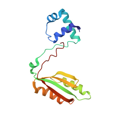
Genome analyses have revealed that members of the Lrp/AsnC family of transcriptional regulators are widely distributed among prokaryotes, including both bacteria and archaea. These regulatory proteins are involved in cellular metabolism in both global and specific manners, depending on the availability of the exogenous amino acid effectors. Here we report the first crystal structure of glutamine receptor protein (Grp) from Sulfolobus tokodaii strain 7, in the ligand-free and glutamine-bound (Grp-Gln) forms. Although the overall structures of both molecules are similar, a significant conformational change was observed at the ligand [L-glutamine (Gln)] binding site in the effector domain, which may be essential for further stabilization of the octameric structure, and in turn for facilitating DNA binding. In addition, we predicted promoter for the grp gene, and these analyses suggested the importance of cooperative binding to the protein. To gain insights into the ligand-induced conformational changes, we mutated all of the ligand-binding residues in Grp, and revealed the importance of Gln binding by biochemical and structural analyses. Further structural analyses showed that Y77 is crucial for ligand binding, and that the residues T132 and T134, which are highly conserved among the Lrp family of proteins, fluctuates between the active and inactive conformations, thus affecting protein oligomerization for DNA binding.
- RIKEN SPring-8 Center, Harima Institute, 1-1-1 Kouto, Sayo, Hyogo 679-5148, Japan. tskvel@spring8.or.jp
Organizational Affiliation: