2PJW | pdb_00002pjw

The Vps27/Hse1 complex is a GAT domain-based scaffold for ubiquitin-dependent sorting


Experimental Data Snapshot

  • Method: X-RAY DIFFRACTION
  • Resolution: 3.01 Å
  • R-Value Free: 
    0.285 (Depositor), 0.291 (DCC) 
  • R-Value Work: 
    0.222 (Depositor), 0.233 (DCC) 
  • R-Value Observed: 
    0.222 (Depositor) 

wwPDB Validation 3D Report Full Report

Validation slider image for 2PJW

This is version 1.4 of the entry. See complete history

Literature

The Vps27/Hse1 Complex Is a GAT Domain-Based Scaffold for Ubiquitin-Dependent Sorting.

Prag, G.Watson, H.Kim, Y.C.Beach, B.M.Ghirlando, R.Hummer, G.Bonifacino, J.S.Hurley, J.H.

(2007) Dev Cell 12: 973-986

  • DOI: https://doi.org/10.1016/j.devcel.2007.04.013
  • Primary Citation Related Structures: 
    2PJW

  • PubMed Abstract: 

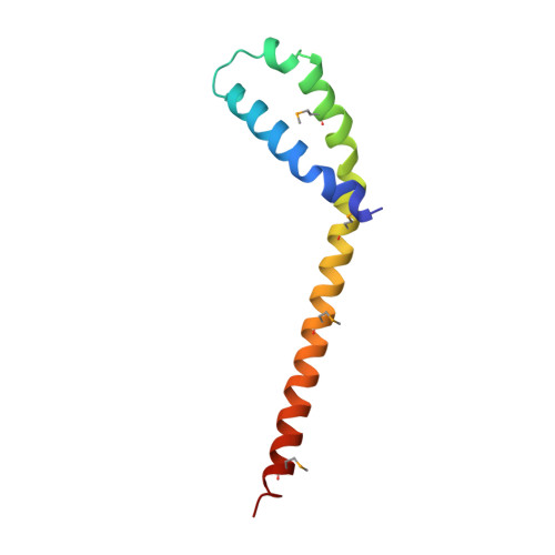
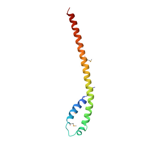
    The yeast Vps27/Hse1 complex and the homologous mammalian Hrs/STAM complex deliver ubiquitinated transmembrane proteins to the ESCRT endosomal-sorting pathway. The Vps27/Hse1 complex directly binds to ubiquitinated transmembrane proteins and recruits both ubiquitin ligases and deubiquitinating enzymes. We have solved the crystal structure of the core responsible for the assembly of the Vps27/Hse1 complex at 3.0 A resolution. The structure consists of two intertwined GAT domains, each consisting of two helices from one subunit and one from the other. The two GAT domains are connected by an antiparallel coiled coil, forming a 90 A-long barbell-like structure. This structure places the domains of Vps27 and Hse1 that recruit ubiquitinated cargo and deubiquitinating enzymes close to each other. Coarse-grained Monte Carlo simulations of the Vps27/Hse1 complex on a membrane show how the complex binds cooperatively to lipids and ubiquitinated membrane proteins and acts as a scaffold for ubiquitination reactions.


  • Organizational Affiliation
    • Laboratory of Molecular Biology, National Institute of Diabetes and Digestive and Kidney Diseases, National Institutes of Health, U.S. Department of Health and Human Services, Bethesda, MD 20892, USA.

Macromolecule Content 

  • Total Structure Weight: 20.82 kDa 
  • Atom Count: 1,458 
  • Modeled Residue Count: 179 
  • Deposited Residue Count: 179 
  • Unique protein chains: 2

Macromolecules

Find similar proteins by:|  3D Structure
Entity ID: 1
MoleculeChains  Sequence LengthOrganismDetailsImage
Uncharacterized protein YHL002WA [auth H]88Saccharomyces cerevisiaeMutation(s): 4 
Gene Names: hse1
UniProt
Find proteins for P38753 (Saccharomyces cerevisiae (strain ATCC 204508 / S288c))
Explore P38753 
Go to UniProtKB:  P38753
Entity Groups
Sequence Clusters30% Identity50% Identity70% Identity90% Identity95% Identity100% Identity
UniProt GroupP38753
Sequence Annotations
Expand
Reference Sequence
Find similar proteins by:|  3D Structure
Entity ID: 2
MoleculeChains  Sequence LengthOrganismDetailsImage
Vacuolar protein sorting-associated protein 27B [auth V]91Saccharomyces cerevisiaeMutation(s): 4 
Gene Names: VPS27GRD11
UniProt
Find proteins for P40343 (Saccharomyces cerevisiae (strain ATCC 204508 / S288c))
Explore P40343 
Go to UniProtKB:  P40343
Entity Groups
Sequence Clusters30% Identity50% Identity70% Identity90% Identity95% Identity100% Identity
UniProt GroupP40343
Sequence Annotations
Expand
Reference Sequence

Small Molecules

Modified Residues  1 Unique
IDChains TypeFormula2D DiagramParent
MSE
Query on MSE
A [auth H]L-PEPTIDE LINKINGC5 H11 N O2 SeMET

Experimental Data & Validation

Experimental Data

  • Method: X-RAY DIFFRACTION
  • Resolution: 3.01 Å
  • R-Value Free:  0.285 (Depositor), 0.291 (DCC) 
  • R-Value Work:  0.222 (Depositor), 0.233 (DCC) 
  • R-Value Observed: 0.222 (Depositor) 
Space Group: P 32 2 1
Unit Cell:
Length ( Å )Angle ( ˚ )
a = 62.4α = 90
b = 62.4β = 90
c = 94.56γ = 120
Software Package:
Software NamePurpose
CNSrefinement
HKL-2000data collection
DENZOdata reduction
HKL-2000data scaling
SnBphasing

Structure Validation

View Full Validation Report


Entry History 

Deposition Data

Revision History  (Full details and data files)

  • Version 1.0: 2007-06-05
    Type: Initial release
  • Version 1.1: 2008-05-01
    Changes: Version format compliance
  • Version 1.2: 2011-07-13
    Changes: Version format compliance
  • Version 1.3: 2017-10-18
    Changes: Refinement description
  • Version 1.4: 2024-11-13
    Changes: Data collection, Database references, Derived calculations, Structure summary