Suprafacial orientation of the SCFCdc4 dimer accommodates multiple geometries for substrate ubiquitination.
Tang, X., Orlicky, S., Lin, Z., Willems, A., Neculai, D., Ceccarelli, D., Mercurio, F., Shilton, B.H., Sicheri, F., Tyers, M.(2007) Cell 129: 1165-1176
- PubMed: 17574027 Search on PubMed
- DOI: https://doi.org/10.1016/j.cell.2007.04.042
- Primary Citation Related Structures:
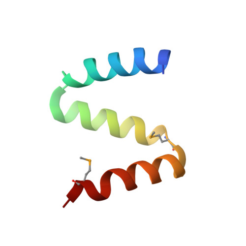
2P63, 2P64 - PubMed Abstract:
SCF ubiquitin ligases recruit substrates for degradation via F box protein adaptor subunits. WD40 repeat F box proteins, such as Cdc4 and beta-TrCP, contain a conserved dimerization motif called the D domain. Here, we report that the D domain protomers of yeast Cdc4 and human beta-TrCP form a superhelical homotypic dimer. Disruption of the D domain compromises the activity of yeast SCF(Cdc4) toward the CDK inhibitor Sic1 and other substrates. SCF(Cdc4) dimerization has little effect on the affinity for Sic1 but markedly stimulates ubiquitin conjugation. A model of the dimeric holo-SCF(Cdc4) complex based on small-angle X-ray scatter measurements reveals a suprafacial configuration, in which substrate-binding sites and E2 catalytic sites lie in the same plane with a separation of 64 A within and 102 A between each SCF monomer. This spatial variability may accommodate diverse acceptor lysine geometries in both substrates and the elongating ubiquitin chain and thereby increase catalytic efficiency.
- Centre for Systems Biology, Samuel Lunenfeld Research Institute, Toronto, Ontario, Canada M5G 1X5.
Organizational Affiliation: