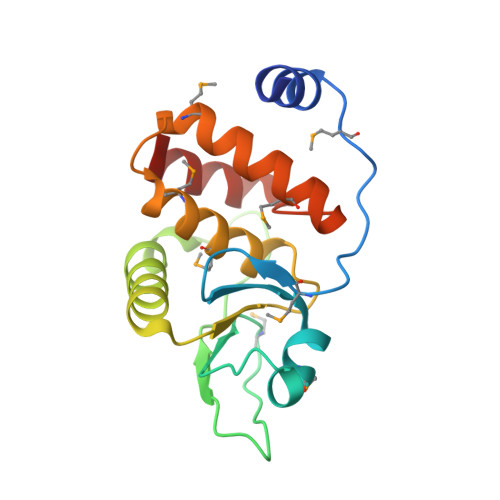
2P4D

Structure-assisted discovery of Variola major H1 phosphatase inhibitors


Experimental Data Snapshot

  • Method: X-RAY DIFFRACTION
  • Resolution: 1.80 Å
  • R-Value Free: 0.195 
  • R-Value Work: 0.178 
  • R-Value Observed: 0.179 

wwPDB Validation   3D Report Full Report


This is version 1.3 of the entry. See complete history


Literature

Structure-assisted discovery of variola major H1 phosphatase inhibitors

Phan, J.Tropea, J.E.Waugh, D.S.

(2007) Acta Crystallogr D Biol Crystallogr D63: 698-704

  • DOI: https://doi.org/10.1107/S0907444907014904
  • Primary Citation of Related Structures:  
    2P4D

  • PubMed Abstract: 

    Variola major virus, the causative agent of smallpox, encodes the dual-specificity H1 phosphatase. Because this enzyme is essential for the production of mature virus particles, it is an attractive molecular target for the development of therapeutic countermeasures for this potential agent of bioterrorism. As a first step in this direction, the crystal structure of H1 phosphatase has been determined at a resolution of 1.8 A. In silico screening methods have led to the identification of several small molecules that inhibit Variola H1 phosphatase with IC(50) values in the low micromolar range. These molecules provide novel leads for future drug development.


  • Organizational Affiliation

    Macromolecular Crystallography Laboratory, Center for Cancer Research, National Cancer Institute at Frederick, PO Box B, Frederick, MD, USA.


Macromolecules
Find similar proteins by:  (by identity cutoff)  |  3D Structure
Entity ID: 1
MoleculeChains Sequence LengthOrganismDetailsImage
Dual specificity protein phosphatase172Variola virusMutation(s): 8 
EC: 3.1.3.48 (PDB Primary Data), 3.1.3.16 (PDB Primary Data)
Entity Groups  
Sequence Clusters30% Identity50% Identity70% Identity90% Identity95% Identity100% Identity
Sequence Annotations
Expand
  • Reference Sequence
Small Molecules
Modified Residues  1 Unique
IDChains TypeFormula2D DiagramParent
MSE
Query on MSE
A
L-PEPTIDE LINKINGC5 H11 N O2 SeMET
Experimental Data & Validation

Experimental Data

  • Method: X-RAY DIFFRACTION
  • Resolution: 1.80 Å
  • R-Value Free: 0.195 
  • R-Value Work: 0.178 
  • R-Value Observed: 0.179 
  • Space Group: I 41 2 2
Unit Cell:
Length ( Å )Angle ( ˚ )
a = 101.441α = 90
b = 101.441β = 90
c = 95.101γ = 90
Software Package:
Software NamePurpose
REFMACrefinement
PDB_EXTRACTdata extraction
MAR345data collection
HKL-2000data reduction
HKL-2000data scaling
SHARPphasing

Structure Validation

View Full Validation Report


Entry History 

Deposition Data

Revision History  (Full details and data files)

  • Version 1.0: 2007-05-29
    Type: Initial release
  • Version 1.1: 2008-05-01
    Changes: Version format compliance
  • Version 1.2: 2011-07-13
    Changes: Version format compliance
  • Version 1.3: 2017-10-11
    Changes: Data collection, Refinement description