2OXD | pdb_00002oxd

Protein kinase CK2 in complex with tetrabromobenzoimidazole K17, K22 and K32 inhibitors


Experimental Data Snapshot

  • Method: X-RAY DIFFRACTION
  • Resolution: 2.30 Å
  • R-Value Free: 
    0.273 (Depositor), 0.266 (DCC) 
  • R-Value Work: 
    0.221 (Depositor), 0.216 (DCC) 
  • R-Value Observed: 
    0.223 (Depositor) 

Starting Model: experimental
View more details

wwPDB Validation 3D Report Full Report

Validation slider image for 2OXD

Ligand Structure Quality Assessment 


This is version 1.2 of the entry. See complete history

Literature

The ATP-Binding Site of Protein Kinase CK2 Holds a Positive Electrostatic Area and Conserved Water Molecules.

Battistutta, R.Mazzorana, M.Cendron, L.Bortolato, A.Sarno, S.Kazimierczuk, Z.Zanotti, G.Moro, S.Pinna, L.A.

(2007) Chembiochem 8: 1804-1809

  • DOI: https://doi.org/10.1002/cbic.200700307
  • Primary Citation Related Structures: 
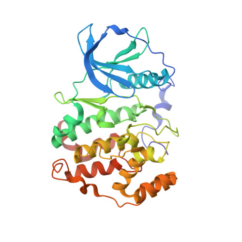
    2OXD, 2OXX, 2OXY

  • PubMed Abstract: 

    CK2 is a highly pleiotropic Ser/Thr protein kinase that is able to promote cell survival and enhance the tumour phenotype under specific circumstances. We have determined the crystal structure of three new complexes with tetrabromobenzimidazole derivatives that display K(i) values between 0.15 and 0.30 microM. A comparative analysis of these data with those of four other inhibitors of the same family revealed the presence of some highly conserved water molecules in the ATP-binding site. These waters reside near Lys68, in an area with a positive electrostatic potential that is able to attract and orient negatively charged ligands. The presence of this positive region and two unique bulky residues that are typical of CK2, Ile66 and Ile174, play a critical role in determining the ligand orientation and binding selectivity.


  • Organizational Affiliation
    • Department of Chemical Sciences, University of Padova, Via Marzolo 1, 35131 Padova, Italy. roberto. battistutta@unipd.it

Macromolecule Content 

  • Total Structure Weight: 39.74 kDa 
  • Atom Count: 2,806 
  • Modeled Residue Count: 325 
  • Deposited Residue Count: 332 
  • Unique protein chains: 1

Macromolecules

Find similar proteins by:|  3D Structure
Entity ID: 1
MoleculeChains  Sequence LengthOrganismDetailsImage
Casein kinase II subunit alpha332Zea maysMutation(s): 0 
Gene Names: ACK2
EC: 2.7.11.1
UniProt
Find proteins for P28523 (Zea mays)
Explore P28523 
Go to UniProtKB:  P28523
Entity Groups
Sequence Clusters30% Identity50% Identity70% Identity90% Identity95% Identity100% Identity
UniProt GroupP28523
Sequence Annotations
Expand
Reference Sequence

Small Molecules

Ligands 1 Unique
IDChains Name / Formula / InChI Key2D Diagram3D Interactions
K32

Query on K32


Download:Ideal Coordinates CCD File
B [auth A]4,5,6,7-TETRABROMO-1H,3H-BENZIMIDAZOL-2-ONE
C7 H2 Br4 N2 O
BMVNFJCUHHTZQU-UHFFFAOYSA-N

Experimental Data & Validation

Experimental Data

  • Method: X-RAY DIFFRACTION
  • Resolution: 2.30 Å
  • R-Value Free:  0.273 (Depositor), 0.266 (DCC) 
  • R-Value Work:  0.221 (Depositor), 0.216 (DCC) 
  • R-Value Observed: 0.223 (Depositor) 
Space Group: C 1 2 1
Unit Cell:
Length ( Å )Angle ( ˚ )
a = 142.148α = 90
b = 60.316β = 103.56
c = 46.238γ = 90
Software Package:
Software NamePurpose
REFMACrefinement
MOSFLMdata reduction
SCALAdata scaling
AMoREphasing

Structure Validation

View Full Validation Report


Ligand Structure Quality Assessment 


Entry History 

Deposition Data

Revision History  (Full details and data files)

  • Version 1.0: 2007-09-25
    Type: Initial release
  • Version 1.1: 2011-07-13
    Changes: Version format compliance
  • Version 1.2: 2023-08-30
    Changes: Data collection, Database references, Derived calculations, Refinement description