2OST | pdb_00002ost

The structure of a bacterial homing endonuclease : I-Ssp6803I


Experimental Data Snapshot

  • Method: X-RAY DIFFRACTION
  • Resolution: 3.10 Å
  • R-Value Free: 
    0.328 (Depositor), 0.302 (DCC) 
  • R-Value Work: 
    0.278 (Depositor), 0.261 (DCC) 
  • R-Value Observed: 
    0.281 (Depositor) 

wwPDB Validation 3D Report Full Report

Validation slider image for 2OST

This is version 1.5 of the entry. See complete history

Literature

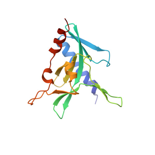
The restriction fold turns to the dark side: a bacterial homing endonuclease with a PD-(D/E)-XK motif.

Zhao, L.Bonocora, R.P.Shub, D.A.Stoddard, B.L.

(2007) EMBO J 26: 2432-2442

  • DOI: https://doi.org/10.1038/sj.emboj.7601672
  • Primary Citation Related Structures: 
    2OST

  • PubMed Abstract: 

    The homing endonuclease I-Ssp6803I causes the insertion of a group I intron into a bacterial tRNA gene-the only example of an invasive mobile intron within a bacterial genome. Using a computational fold prediction, mutagenic screen and crystal structure determination, we demonstrate that this protein is a tetrameric PD-(D/E)-XK endonuclease - a fold normally used to protect a bacterial genome from invading DNA through the action of restriction endonucleases. I-Ssp6803I uses its tetrameric assembly to promote recognition of a single long target site, whereas restriction endonuclease tetramers facilitate cooperative binding and cleavage of two short sites. The limited use of the PD-(D/E)-XK nucleases by mobile introns stands in contrast to their frequent use of LAGLIDADG and HNH endonucleases - which in turn, are rarely incorporated into restriction/modification systems.


  • Organizational Affiliation
    • Graduate Program in Molecular Biophysics, Structure and Design, University of Washington, Seattle, WA, USA.

Macromolecule Content 

  • Total Structure Weight: 87.53 kDa 
  • Atom Count: 5,747 
  • Modeled Residue Count: 625 
  • Deposited Residue Count: 662 
  • Unique protein chains: 1
  • Unique nucleic acid chains: 2

Macromolecules


Find similar proteins by:|  3D Structure
Entity ID: 3
MoleculeChains  Sequence LengthOrganismDetailsImage
Putative endonucleaseC [auth A],
D [auth B],
E [auth C],
F [auth D]
151Synechocystis sp. PCC 6803Mutation(s): 4 
UniProt
Find proteins for Q57253 (Synechocystis sp. (strain ATCC 27184 / PCC 6803 / Kazusa))
Explore Q57253 
Go to UniProtKB:  Q57253
Entity Groups
Sequence Clusters30% Identity50% Identity70% Identity90% Identity95% Identity100% Identity
UniProt GroupQ57253
Sequence Annotations
Expand
Reference Sequence
Find similar nucleic acids by:  (by identity cutoff) 
Entity ID: 1
MoleculeChains LengthOrganismImage
Synthetic DNA 29 MERA [auth Y]29N/A
Sequence Annotations
Expand
Reference Sequence
Find similar nucleic acids by:  (by identity cutoff) 
Entity ID: 2
MoleculeChains LengthOrganismImage
Synthetic DNA 29 MERB [auth Z]29N/A
Sequence Annotations
Expand
Reference Sequence

Experimental Data & Validation

Experimental Data

  • Method: X-RAY DIFFRACTION
  • Resolution: 3.10 Å
  • R-Value Free:  0.328 (Depositor), 0.302 (DCC) 
  • R-Value Work:  0.278 (Depositor), 0.261 (DCC) 
  • R-Value Observed: 0.281 (Depositor) 
Space Group: I 4 2 2
Unit Cell:
Length ( Å )Angle ( ˚ )
a = 143.777α = 90
b = 143.777β = 90
c = 319.179γ = 90
Software Package:
Software NamePurpose
DENZOdata reduction
SCALEPACKdata scaling
SHARPphasing
DMphasing
REFMACrefinement
PDB_EXTRACTdata extraction
ADSCdata collection

Structure Validation

View Full Validation Report


Entry History 

Deposition Data

Revision History  (Full details and data files)

  • Version 1.0: 2007-03-20
    Type: Initial release
  • Version 1.1: 2008-05-01
    Changes: Version format compliance
  • Version 1.2: 2011-07-13
    Changes: Source and taxonomy, Version format compliance
  • Version 1.3: 2017-10-18
    Changes: Refinement description
  • Version 1.4: 2021-10-20
    Changes: Database references, Derived calculations
  • Version 1.5: 2024-02-21
    Changes: Data collection, Refinement description