An alternate description of two crystal structures of phospholipase A(2) from Bungarus caeruleus.
Le Trong, I., Stenkamp, R.E.(2007) Acta Crystallogr D Biol Crystallogr 63: 548-549
- PubMed: 17372360 Search on PubMed
- DOI: https://doi.org/10.1107/S0907444907007354
- Primary Citation Related Structures:
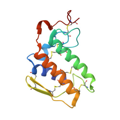
2OSN - PubMed Abstract:
Reinterpretations of the space-group symmetry are reported for two crystal structures of phospholipase A(2) isoforms (PDB codes 1u4j and 1g2x). The two structures reported in space groups R3 and C2 are isomorphous with a third isoform with space group R32 (PDB code 1fe5). The original structure reports were interpreted in terms of different oligomeric forms of the isoforms, but these conclusions are not supported by the isomorphous structures.
- Departments of Biological Structure and Biochemistry, Biomolecular Structure Center, University of Washington, Seattle, WA 98195, USA.
Organizational Affiliation: