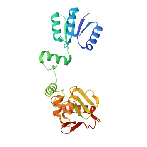
2OQR

The structure of the response regulator RegX3 from Mycobacterium tuberculosis


Experimental Data Snapshot

  • Method: X-RAY DIFFRACTION
  • Resolution: 2.03 Å
  • R-Value Free: 0.217 
  • R-Value Work: 0.177 
  • R-Value Observed: 0.179 

wwPDB Validation   3D Report Full Report


This is version 1.3 of the entry. See complete history


Literature

The Structure of a Full-length Response Regulator from Mycobacterium tuberculosis in a Stabilized Three-dimensional Domain-swapped, Activated State.

King-Scott, J.Nowak, E.Mylonas, E.Panjikar, S.Roessle, M.Svergun, D.I.Tucker, P.A.

(2007) J Biol Chem 282: 37717-37729

  • DOI: https://doi.org/10.1074/jbc.M705081200
  • Primary Citation of Related Structures:  
    2OQR

  • PubMed Abstract: 

    The full-length, two-domain response regulator RegX3 from Mycobacterium tuberculosis is a dimer stabilized by three-dimensional domain swapping. Dimerization is known to occur in the OmpR/PhoB subfamily of response regulators upon activation but has previously only been structurally characterized for isolated receiver domains. The RegX3 dimer has a bipartite intermolecular interface, which buries 2357 A(2) per monomer. The two parts of the interface are between the two receiver domains (dimerization interface) and between a composite receiver domain and the effector domain of the second molecule (interdomain interface). The structure provides support for the importance of threonine and tyrosine residues in the signal transduction mechanism. These residues occur in an active-like conformation stabilized by lanthanum ions. In solution, RegX3 exists as both a monomer and a dimer in a concentration-dependent equilibrium. The dimer in solution differs from the active form observed in the crystal, resembling instead the model of the inactive full-length response regulator PhoB.


  • Organizational Affiliation

    EMBL-Hamburg Outstation, c/o DESY, Notkestrasse 85, D-22603, Hamburg, Germany.


Macromolecules
Find similar proteins by:  (by identity cutoff)  |  3D Structure
Entity ID: 1
MoleculeChains Sequence LengthOrganismDetailsImage
Sensory transduction protein regX3230Mycobacterium tuberculosis H37RvMutation(s): 0 
Gene Names: regX3
UniProt
Find proteins for P9WGL9 (Mycobacterium tuberculosis (strain ATCC 25618 / H37Rv))
Explore P9WGL9 
Go to UniProtKB:  P9WGL9
Entity Groups  
Sequence Clusters30% Identity50% Identity70% Identity90% Identity95% Identity100% Identity
UniProt GroupP9WGL9
Sequence Annotations
Expand
  • Reference Sequence
Small Molecules
Ligands 3 Unique
IDChains Name / Formula / InChI Key2D Diagram3D Interactions
LA
Query on LA

Download Ideal Coordinates CCD File 
D [auth A],
E [auth A],
F [auth A],
G [auth A],
H [auth A]
LANTHANUM (III) ION
La
CZMAIROVPAYCMU-UHFFFAOYSA-N
BME
Query on BME

Download Ideal Coordinates CCD File 
I [auth A]BETA-MERCAPTOETHANOL
C2 H6 O S
DGVVWUTYPXICAM-UHFFFAOYSA-N
ACT
Query on ACT

Download Ideal Coordinates CCD File 
B [auth A],
C [auth A]
ACETATE ION
C2 H3 O2
QTBSBXVTEAMEQO-UHFFFAOYSA-M
Experimental Data & Validation

Experimental Data

  • Method: X-RAY DIFFRACTION
  • Resolution: 2.03 Å
  • R-Value Free: 0.217 
  • R-Value Work: 0.177 
  • R-Value Observed: 0.179 
  • Space Group: P 4 21 2
Unit Cell:
Length ( Å )Angle ( ˚ )
a = 124.351α = 90
b = 124.351β = 90
c = 44.824γ = 90
Software Package:
Software NamePurpose
DENZOdata reduction
SCALEPACKdata scaling
REFMACrefinement
PDB_EXTRACTdata extraction
Auto-Rickshawphasing

Structure Validation

View Full Validation Report


Entry History 

Deposition Data

Revision History  (Full details and data files)

  • Version 1.0: 2007-10-16
    Type: Initial release
  • Version 1.1: 2011-07-13
    Changes: Derived calculations, Version format compliance
  • Version 1.2: 2017-10-18
    Changes: Refinement description
  • Version 1.3: 2023-12-27
    Changes: Data collection, Database references, Derived calculations