2OO5

Structure of transhydrogenase (dI.H2NADH)2(dIII.NADP+)1 asymmetric complex


Experimental Data Snapshot

  • Method: X-RAY DIFFRACTION
  • Resolution: 2.60 Å
  • R-Value Free: 0.275 
  • R-Value Work: 0.219 
  • R-Value Observed: 0.222 

wwPDB Validation   3D Report Full Report


Ligand Structure Quality Assessment 


This is version 1.3 of the entry. See complete history


Literature

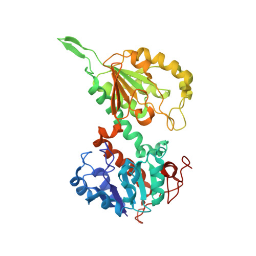
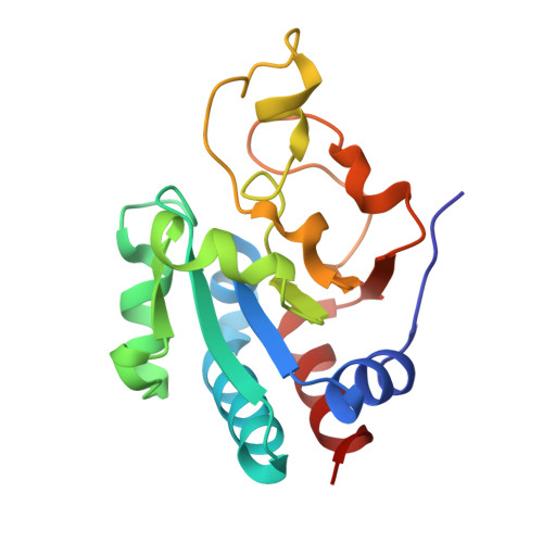
Structures of the dI(2)dIII(1) Complex of Proton-Translocating Transhydrogenase with Bound, Inactive Analogues of NADH and NADPH Reveal Active Site Geometries

Bhakta, T.Whitehead, S.J.Snaith, J.S.Dafforn, T.R.Wilkie, J.Rajesh, S.White, S.A.Jackson, J.B.

(2007) Biochemistry 46: 3304-3318

  • DOI: https://doi.org/10.1021/bi061843r
  • Primary Citation of Related Structures:  
    2OO5, 2OOR

  • PubMed Abstract: 

    Transhydrogenase couples the redox reaction between NADH and NADP+ to proton translocation across a membrane. The enzyme comprises three components; dI binds NAD(H), dIII binds NADP(H), and dII spans the membrane. The 1,4,5,6-tetrahydro analogue of NADH (designated H2NADH) bound to isolated dI from Rhodospirillum rubrum transhydrogenase with similar affinity to the physiological nucleotide. Binding of either NADH or H2NADH led to closure of the dI mobile loop. The 1,4,5,6-tetrahydro analogue of NADPH (H2NADPH) bound very tightly to isolated R. rubrum dIII, but the rate constant for dissociation was greater than that for NADPH. The replacement of NADP+ on dIII either with H2NADPH or with NADPH caused a similar set of chemical shift alterations, signifying an equivalent conformational change. Despite similar binding properties to the natural nucleotides, neither H2NADH nor H2NADPH could serve as a hydride donor in transhydrogenation reactions. Mixtures of dI and dIII form dI2dIII1 complexes. The nucleotide charge distribution of complexes loaded either with H2NADH and NADP+ or with NAD+ and H2NADPH should more closely mimic the ground states for forward and reverse hydride transfer, respectively, than previously studied dead-end species. Crystal structures of such complexes at 2.6 and 2.3 A resolution are described. A transition state for hydride transfer between dihydronicotinamide and nicotinamide derivatives determined in ab initio quantum mechanical calculations resembles the organization of nucleotides in the transhydrogenase active site in the crystal structure. Molecular dynamics simulations of the enzyme indicate that the (dihydro)nicotinamide rings remain close to a ground state for hydride transfer throughout a 1.4 ns trajectory.


  • Organizational Affiliation

    School of Biosciences, University of Birmingham, Edgbaston, UK.


Macromolecules
Find similar proteins by:  (by identity cutoff)  |  3D Structure
Entity ID: 1
MoleculeChains Sequence LengthOrganismDetailsImage
NAD(P) transhydrogenase subunit alpha part 1
A, B
384Rhodospirillum rubrumMutation(s): 0 
Gene Names: pntAAnntA1
EC: 1.6.1.2
UniProt
Find proteins for Q2RSB2 (Rhodospirillum rubrum (strain ATCC 11170 / ATH 1.1.1 / DSM 467 / LMG 4362 / NCIMB 8255 / S1))
Explore Q2RSB2 
Go to UniProtKB:  Q2RSB2
Entity Groups  
Sequence Clusters30% Identity50% Identity70% Identity90% Identity95% Identity100% Identity
UniProt GroupQ2RSB2
Sequence Annotations
Expand
  • Reference Sequence
Find similar proteins by:  (by identity cutoff)  |  3D Structure
Entity ID: 2
MoleculeChains Sequence LengthOrganismDetailsImage
NAD(P) transhydrogenase subunit beta174Rhodospirillum rubrumMutation(s): 0 
Gene Names: pntBnntB
EC: 1.6.1.2
UniProt
Find proteins for P0C188 (Rhodospirillum rubrum)
Explore P0C188 
Go to UniProtKB:  P0C188
Entity Groups  
Sequence Clusters30% Identity50% Identity70% Identity90% Identity95% Identity100% Identity
UniProt GroupP0C188
Sequence Annotations
Expand
  • Reference Sequence
Experimental Data & Validation

Experimental Data

  • Method: X-RAY DIFFRACTION
  • Resolution: 2.60 Å
  • R-Value Free: 0.275 
  • R-Value Work: 0.219 
  • R-Value Observed: 0.222 
  • Space Group: P 21 21 21
Unit Cell:
Length ( Å )Angle ( ˚ )
a = 71.199α = 90
b = 101.025β = 90
c = 131.721γ = 90
Software Package:
Software NamePurpose
SCALAdata scaling
REFMACrefinement
PDB_EXTRACTdata extraction
MOSFLMdata reduction
CCP4data scaling

Structure Validation

View Full Validation Report


Ligand Structure Quality Assessment 


Entry History 

Deposition Data

Revision History  (Full details and data files)

  • Version 1.0: 2007-03-13
    Type: Initial release
  • Version 1.1: 2008-05-01
    Changes: Version format compliance
  • Version 1.2: 2011-07-13
    Changes: Version format compliance
  • Version 1.3: 2023-08-30
    Changes: Data collection, Database references, Derived calculations, Refinement description