2OLW | pdb_00002olw

Crystal Structure of E. coli pseudouridine synthase RluE


Experimental Data Snapshot

  • Method: X-RAY DIFFRACTION
  • Resolution: 1.60 Å
  • R-Value Free: 
    0.255 (Depositor), 0.257 (DCC) 
  • R-Value Work: 
    0.213 (Depositor), 0.215 (DCC) 
  • R-Value Observed: 
    0.215 (Depositor) 

Starting Model: experimental
View more details

wwPDB Validation 3D Report Full Report

Validation slider image for 2OLW

This is version 1.4 of the entry. See complete history

Literature

The Crystal Structure of E. coli rRNA Pseudouridine Synthase RluE.

Pan, H.Ho, J.D.Stroud, R.M.Finer-Moore, J.

(2007) J Mol Biology 367: 1459-1470

  • DOI: https://doi.org/10.1016/j.jmb.2007.01.084
  • Primary Citation Related Structures: 
    2OLW, 2OML

  • PubMed Abstract: 

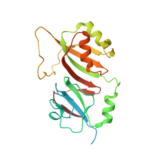
    Pseudouridine synthase RluE modifies U2457 in a stem of 23 S RNA in Escherichia coli. This modification is located in the peptidyl transferase center of the ribosome. We determined the crystal structures of the C-terminal, catalytic domain of E. coli RluE at 1.2 A resolution and of full-length RluE at 1.6 A resolution. The crystals of the full-length enzyme contain two molecules in the asymmetric unit and in both molecules the N-terminal domain is disordered. The protein has an active site cleft, conserved in all other pseudouridine synthases, that contains invariant Asp and Tyr residues implicated in catalysis. An electropositive surface patch that covers the active site cleft is just wide enough to accommodate an RNA stem. The RNA substrate stem can be docked to this surface such that the catalytic Asp is adjacent to the target base, and a conserved Arg is positioned to help flip the target base out of the stem into the enzyme active site. A flexible RluE specific loop lies close to the conserved region of the stem in the model, and may contribute to substrate specificity. The stem alone is not a good RluE substrate, suggesting RluE makes additional interactions with other regions in the ribosome.


  • Organizational Affiliation
    • Department of Biochemistry and Biophysics, University of California at San Francisco, San Francisco, CA 94143, USA.

Macromolecule Content 

  • Total Structure Weight: 50.44 kDa 
  • Atom Count: 3,129 
  • Modeled Residue Count: 358 
  • Deposited Residue Count: 434 
  • Unique protein chains: 1

Macromolecules

Find similar proteins by:|  3D Structure
Entity ID: 1
MoleculeChains  Sequence LengthOrganismDetailsImage
Ribosomal large subunit pseudouridine synthase E
A, B
217Escherichia coliMutation(s): 0 
Gene Names: rluE
EC: 5.4.99 (PDB Primary Data), 5.4.99.20 (UniProt)
UniProt
Find proteins for P75966 (Escherichia coli (strain K12))
Explore P75966 
Go to UniProtKB:  P75966
Entity Groups
Sequence Clusters30% Identity50% Identity70% Identity90% Identity95% Identity100% Identity
UniProt GroupP75966
Sequence Annotations
Expand
Reference Sequence

Small Molecules

Ligands 3 Unique
IDChains Name / Formula / InChI Key2D Diagram3D Interactions
DTT

Query on DTT


Download:Ideal Coordinates CCD File
F [auth B]2,3-DIHYDROXY-1,4-DITHIOBUTANE
C4 H10 O2 S2
VHJLVAABSRFDPM-IMJSIDKUSA-N
SO4

Query on SO4


Download:Ideal Coordinates CCD File
C [auth A]SULFATE ION
O4 S
QAOWNCQODCNURD-UHFFFAOYSA-L
ACY

Query on ACY


Download:Ideal Coordinates CCD File
D [auth A]
E [auth A]
G [auth B]
H [auth B]
I [auth B]
D [auth A],
E [auth A],
G [auth B],
H [auth B],
I [auth B],
J [auth B]
ACETIC ACID
C2 H4 O2
QTBSBXVTEAMEQO-UHFFFAOYSA-N

Experimental Data & Validation

Experimental Data

  • Method: X-RAY DIFFRACTION
  • Resolution: 1.60 Å
  • R-Value Free:  0.255 (Depositor), 0.257 (DCC) 
  • R-Value Work:  0.213 (Depositor), 0.215 (DCC) 
  • R-Value Observed: 0.215 (Depositor) 
Space Group: P 21 21 21
Unit Cell:
Length ( Å )Angle ( ˚ )
a = 59.015α = 90
b = 78.887β = 90
c = 84.182γ = 90
Software Package:
Software NamePurpose
DENZOdata reduction
SCALEPACKdata scaling
REFMACrefinement
PDB_EXTRACTdata extraction
ELVESrefinement
CNSphasing

Structure Validation

View Full Validation Report


Entry History 

Deposition Data

Revision History  (Full details and data files)

  • Version 1.0: 2007-03-13
    Type: Initial release
  • Version 1.1: 2008-05-01
    Changes: Version format compliance
  • Version 1.2: 2011-07-13
    Changes: Version format compliance
  • Version 1.3: 2017-10-18
    Changes: Refinement description
  • Version 1.4: 2023-08-30
    Changes: Data collection, Database references, Derived calculations, Refinement description